Abstract
Background. In the domain of forensic medicine, the estimation of age is a critical aspect of human identification, including that of adolescents.
Objectives. The study aimed to examine the relationship between the pulp chamber volume of teeth with open apices and the chronological age of adolescents from the Turkish population.
Material and methods. The study was conducted by examining cone beam computed tomography (CBCT) images of 51 pediatric patients who visited the Department of Pedodontics for routine dental examinations. All CBCT images were captured using a NewTom 5G unit (QR, Verona, Italy). The measurements were recorded in DICOM format using the SimPlant Pro 16 software (Materialise NV, Leuven, Belgium).
Results. A moderate and positive significant correlation was observed between the age of the premolar group patients and impacted pulp volume (IMPV). The increase in volume measurements of the patients in the premolar group indicates that their age is significantly higher (r = 0.561, p = 0.030). A moderate and negative significant relationship was identified between the age of the premolar group patients and erupted pulp volume (EPV). The increase in the EPV measurements of the patients in the premolar group indicates that their age is significantly lower (r = –0.491, p = 0.041).
Conclusions. The pulp chamber volume of premolars, which narrows due to secondary dentin deposition, served as the physical marker for chronological age estimation of adolescents in the Turkish population. The measurement of the mature and immature premolar pulp volume could be used for age estimation, particularly in the context of CBCT analysis.
Keywords: cone beam computed tomography, dentin, bicuspid, forensic dentistry, chronological age
Introduction
The estimation of age is a critical aspect of human identification for forensic purposes. When a professional medical opinion is required to determine an individual’s status regarding, for example, adoption or punitive offenses or whether the individual is a juvenile or adult, the estimation of their age can be critical. Due to its frequent application in legal contexts, the determination of age has become increasingly significant in the field of forensic medicine, including adolescent cases.1, 2 However, age estimation can be influenced by developmental changes, as biological age and chronological age are not necessarily aligned.3, 4
The scientific literature has described different methods for assessing age indicators related to skeletal changes.5 In addition, many techniques have been investigated for age-related changes in dental structure, which are less influenced by nutritional and endocrine factors.5, 6 Given that environmental and pathological aspects have a smaller influence on tooth development than skeletal factors,1 particularly during the preadult stage, tooth development is frequently used to determine the age of individuals. This method has been found to offer more accurate results than conventional methods used for adult age estimation.6
Dental age can be determined by radiological, morphophysiological and biochemical methods.7 Radiological techniques have become popular due to their simplicity and reproducibility in applications on both living and deceased individuals.7, 8 Periapical and panoramic radiography along with advanced digital technologies for imaging lateral oblique and cephalometric views, are particularly effective in age identification.8 In general, radiological methods of age estimation are based on the evaluation of properties such as the development and resemblance of tooth germs, the earliest detectable sign or beginning of mineralization, the degree of crown completion, the degree of root completion of erupted or unerupted teeth, the degree of resorption of primary teeth, the measurement of open apices in teeth, the eruption of crowns into the oral cavity, the volume of the pulp chamber, root canals, and the formation of physiological secondary teeth.2, 7, 8 Based on these factors, the estimated age of patients can serve as a crucial aspect of the planning of interceptive orthodontic treatment for children.9
Due to secondary dentin deposition in the wall of the pulp cavity, odontoblasts gradually shrink the coronal pulp chamber and root volume throughout an individual’s life.5, 10 The majority of dental publications have correlated age with the volume of the dental pulp chamber and secondary dentin apposition.2, 10, 11 The volume of the dental pulp chamber can be measured by using radiographic methods, including 2D or 3D radiography.12 Two-dimensional radiographs are used to estimate age; however, they provide a limited visualization of the dental structure owing to superposition and distortion. This type of radiographs represents linear measurements, which are not sufficient to reveal changes in pulp volume related to secondary dentin apposition in 3 dimensions.13 Even so, studies aimed at determining the age of children with open apices in their teeth have involved 2D radiography.9, 11, 14
Dental modeling, developed with 3D cone beam computed tomography (CBCT), is based on the geometric approximation of the various components of a tooth, including the root, the pulp and the crown. Dental modeling with CBCT is a well-established method that produces reliable results and enables the rapid assessment of the pulp volumes of teeth,2 namely by calculating pulp volume using commercial licensed software.6 However, in studies focused on the correlation between chronological age and the pulp–tooth volume ratio, samples have represented all age groups, from infants to older adults, instead of specific ones. Additionally, few studies have employed the pulp–tooth volume ratio in conjunction with CBCT values to determine the dental age of adolescents.15, 16 Against that background, the aim of this study was to propose a CBCT-based method for age estimation that utilizes the pulp chamber volume in adolescents.
Material and methods
This retrospective study was approved by Erciyes University Non-Invasive Clinical Practices Ethics Committee (decision No. 2022/113). The study was conducted in the Department of Oral and Maxillofacial Radiology and the Department of Pediatric Dentistry of the Faculty of Dentistry at Erciyes University, Kayseri, Turkey. Throughout the study, adherence to the principles of the Declaration of Helsinki was maintained. Informed consent forms were obtained from the parents of all patients.
Inclusion and exclusion criteria
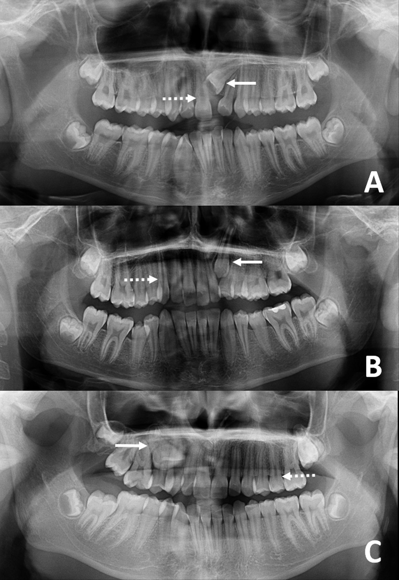
Two radiologists recorded each patient’s sex and chronological age in years and months, while a technician was responsible for taking all images. Firstly, panoramic radiographs were obtained from patients who had applied to our clinic for dental treatment. The CBCT images were taken if necessary, in the presence of impacted teeth or due to other dental reasons (1,598 patients). In order to be included in the study, patients had to be between the ages of 9 and 14 years, as determined by CBCT and panoramic images from 2014–2020, and must have had permanent dentition in the maxillary region. A total of 51 individuals were included in the study. The patients were not requested to undergo radiographic imaging for the study. Panoramic radiography results are depicted in Figure 1.
Patients with caries-affected teeth, teeth with pathological wear, coronal fracture, external and internal resorption, or teeth that had received general dental treatment for odontoma were excluded from the study. Patients with teeth exhibiting abnormal dental anatomy, which complicates measurement, were also excluded.
Pulp volume measurement
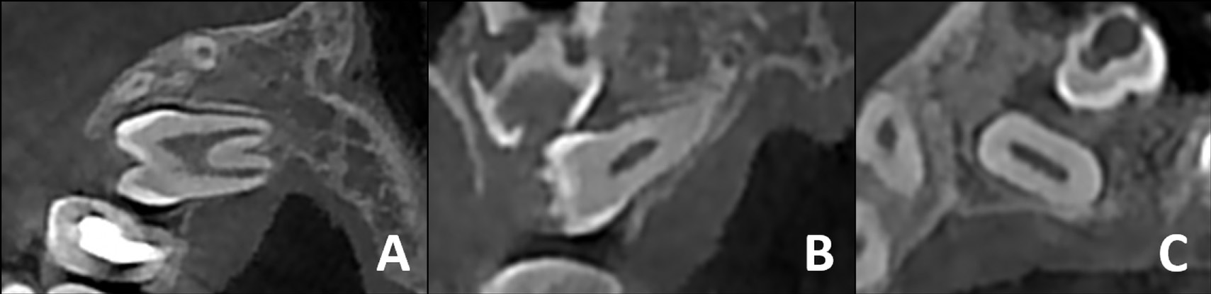
All CBCT images were acquired using a NewTom 5G unit (QR, Verona, Italy) in a standard mode, with a field of view of 12 cm × 8 cm and a voxel size of 250 μm. The kVp and mA values were derived from images obtained through the use of a preview mode. The CBCT images were also examined in terms of the established criteria (Figure 2). Subsequently, a study group was formed.
Radiographs with poor image quality were excluded from the study. The images were analyzed using the Dell Precision T1500 WorkStation (Dell D02M; Dell Technologies, Warsaw, Poland) and a 19-inch 1920 × 1080 pixel resolution Dell monitor (Dell E190S; Dell Technologies, Beijing, China). The reported measurements were recorded in DICOM format using the NNT software, v. 9.01 (NewTom, Verona, Italy) and the NewTom 5G CBCT device, and reconstructed in the SimPlant Pro 16 software (Materialise NV, Leuven, Belgium).
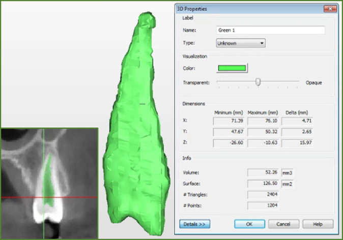
To measure pulp volume from the CBCT data, a threshold for soft tissue value was set, and the mask creation and segmentation technique was used to determine the contour of the pulp and pinpoint the value of the volume. First, a mask was created to form the pulp chamber and hard tissue segments of each involved tooth. Second, the optimal separation grayscale threshold was selected, which exhibited the pulp chamber within the tooth in all sections and planes (i.e., axial, coronal and sagittal). Third, for the 3D calculation, regions that did not correspond to the pulp cavity were approximately identified and deleted. Fourth, manual erasing and corrected drawing were performed to remove bone fragments at the root and in surrounding teeth at the crown level. Standardization was achieved using fixed threshold values in all teeth. Lastly, with the mask representing the pulp chamber in all planes and sections, an image was generated to calculate the pulp volume. The image value was automatically measured by the software, and the pulp volume was obtained (Figure 3).
Statistical analysis
The normality of the data was determined using Q–Q plots, a histogram and the Shapiro–Wilk test. Levene’s test was performed to assess the homogeneity of variance, and a set of descriptive statistics, including frequency, percentage, mean (M), and standard deviation (SD) values, was calculated. In all patients and groups, the relationship between pulp volumes and age was examined using Pearson’s correlation analysis. The multiple regression analysis was utilized to model and clarify the association between age and pulp volume. The analyses were conducted using the IBM SPSS Statistics for Windows software, v. 20.0 (IBM Corp., Armonk, USA). Statistically significant results were defined as p < 0.05.
Results
Of the patients included in the sample, 51% (n = 26) were male, and 49% (n = 25) were female. The patient groups consisted of the incisal group (31.4%), the canine group (37.3%) and the premolar group (31.4%). The mean age of the patients was 145.90 ±18.15 months. The mean impacted dental pulp volume (IMPV) was 67.34 ±21.45 mm3, while the mean erupted pulp volume (EPV) was 56.44 ±18.36 mm3 (Table 1).
No statistically significant difference (p = 0.932, range = 0.889–0.987) was observed between radiologists in pulp measurements. A weak positive correlation was identified between IMPV and age (r = 0.393, p = 0.011), and a weak negative correlation was observed between EPV and age (r = –0.271, p = 0.041) (Table 2).
In the incisal group, no significant correlation was identified between age and IMPV (r = 0.072, p = 0.810), or between age and EPV (r = 0.150, p = 0.594). Likewise, in the canine group, no significant correlation was observed between age and IMPV (r = 0.010, p = 0.993), or between age and EPV (r = –0.162, p = 0.522). However, in the premolar group, there was a moderate positive significant correlation between age and IMPV (r = 0.561, p = 0.032). This finding suggests that as IMPV values increase, the age of the participant is found to be significantly higher. In the same group, there was a moderate negative significant correlation between age and EPV (r = –0.492, p = 0.041), and an elevated EPV value indicated that a subject’s age was significantly lower (Table 3).
In addition, there was a significant correlation between pulp volume and age in the premolar group (p = 0.011). The multiple regression formula for this relationship was calculated as follows (Equation 1):
The model determined that IMPV and EPV in the premolar group affected the age significantly (Table 4).
Discussion
There are distinct differences between the teeth and the palate.17 A digital model of the palate is an excellent candidate for disaster victim identification because it changes only 3 μm per year throughout a person’s life.18 Contrarily, teeth undergo continuous modification. Although teeth are subject to plenty of age-related changes during life, all anatomical dental characteristics are unique, which has made teeth a useful option for comparative identification for centuries.19 However, our results prove that teeth could also be used for age estimation in adolescents. According to the literature, the constriction of pulp dimensions due to secondary deposition can serve as a useful indicator of chronological age.5, 10, 12, 20
Secondary dentin deposition can be assessed using the pulp–tooth volume ratio with 2D dental radiographic methods. However, the primary disadvantage of conventional radiographic techniques is that they are subject to substantial errors in magnification and distortion. Therefore, concurrent evaluation of mesiodistal and buccolingual dimensions of teeth is recommended.19, 20, 21 By contrast, CBCT provides an excellent means to collect high-quality 3D tooth radiographs in dental practice and is more appealing than CT and micro-CT for determining age.22
During the developmental process of a tooth, the diameter of the apical foramen decreases, and the crown develops with enamel production. Cameriere et al. determined the maturity of teeth under these conditions by counting the number of teeth with an entirely closed apical foramen and measuring the distance between the length of the inner point in the apical foramen and the length of the teeth.23 They also quantified the dental age of children by measuring the width of the apical foramen’s opening.23 In most studies, teeth with open apices have been evaluated using 2D radiographic techniques.1, 11, 14, 23, 24 However, with the advancement of medical technology, CBCT has become a valuable tool in dentistry clinics, offering a higher metric resolution and isotropic voxel resolution than conventional medical computed tomography.25 Cone beam computed tomography image analysis using the segmentation function can help to design the scanned structure’s 3D model and ascertain its volume and superficial area.26 Once the pulp–tooth ratio is measured, mature teeth can be evaluated with CBCT.21, 27, 28, 29, 30 In the present study, the authors used CBCT to evaluate both mature and immature teeth.
Researchers have traditionally used incisors, canines and premolars to estimate age, either in isolation or together.15, 21, 22, 27, 29, 30, 31 For example, Star et al. analyzed mono-radicular incisal, canine and premolar teeth in subjects from Belgium aged 8–19 years.27 The authors have used the SimPlant Pro software to calculate pulp volume from 111 CBCT images of teeth. As a result, no statistically significant relationship has been identified between pulp volume and age across different tooth types (p = 0.15), and pulp volume–tooth ratios ranged between 0.002 and 0.091 (0.027 ±0.020). Upon calculating regression formulas for each tooth, it was observed that the relationship between the pulp volume ratio of incisors and age was stronger for females than for males. However, the calculated difference was not statistically significant (p = 0.86), and no significant interaction between types of teeth and sex was observed (p = 0.50).27 Many previous studies have also shown that no significant relationship exists between pulp volume and sex,21, 22, 27, 32, 33 as confirmed by the findings of the present study.
Gulsahi et al. performed a CBCT analysis on a sample of 655 maxillary central incisors, lateral canines, mandibular canines, and first and second premolars in Turkey.21 Following the implementation of a simple linear regression analysis and the utilization of the 3D-DOCTOR software for tissue segmentation, the regression analysis showed that 53.2% of the variance in maxillary central incisors could be explained, along with 21.7%, 21.0%, 20.7%, and 15.3% explanations for the variance observed in mandibular second premolars, mandibular canines, mandibular first premolars, and maxillary canines, respectively.21 In another study, Aboshi et al. investigated the relationship between age and different levels of pulp volume in lower premolars.12 This tooth type was selected due to its superior resistance to decay compared to incisors and canines, and its more straightforward and stable root shape compared to molars.12 The study findings revealed that pulp volume in lower premolars diminished progressively over time, with the largest decline occurring at ages of 20 and 50, and the most substantial loss observed at the coronal third of the root. In addition, the study’s accuracy in estimating age using the model for lower second premolars (R2 = 0.685) exhibited a marginal improvement over that for lower first premolars (R2 = 0.617).12 By comparison, Tardivo et al. examined 210 CT scans from individuals aged 15–85 years with 4 healthy canines.19 The samples composed of 840 canines were modeled using the Mimics® 10.01 software. The authors formulated 7 regression models and determined the most powerful one, which took maxillary canines into consideration.19 The decision to study canines was made due to their relatively simple anatomy and large dimensions of their various volumes.19
This study was performed using the SimPlant Pro 16 software (Materialise NV) with CBCT, and included participants aged 9–14 years. The authors evaluated incisors, canines and premolars collectively to examine the relationship between each tooth and age. A weak positive significant relationship was observed between the age of the patients and IMPV. Therefore, we propose that the rise in IMPV values may signify a considerably higher age of the patients (r = 0.393, p = 0.010). As IMPV volume increases, chronological age increases. Meanwhile, taking into consideration the weak negative significant relationship between the age of the patients and EPV, the increase in EPV values may be indicative of the patients’ significantly lower age (r = –0.271, p = 0.040).
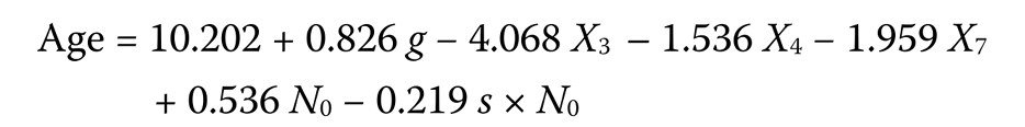
In other comparable studies, Yang et al. examined left maxillary incisors and canines in a sample of individuals aged 8–19 years from China.15 They reported means and standard deviations of the pulp–tooth volume ratio to be 0.053 ±0.024 for male left maxillary incisors, 0.049 ±0.069 for female left maxillary incisors, 0.080 ±0.030 for male left maxillary canines, and 0.020 ±0.022 for female left maxillary canines. The researchers also determined that the correlation coefficients were –0.70 (male), –0.63 (female) and –0.67 (total) for central incisors, and –0.88 (male), –0.81 (female) and –0.83 (total) for canines.15 Meanwhile, Pinchi et al. examined left maxillary central incisors in a sample of Italian individuals aged 10–80 years.22 The authors found that pulp volume was a statistically significant predictor of age (p < 0.001) and calculated the linear regression formula (–64.14 – 32.00 × pulp volume (mm3)) as a reliable parameter for determining age in adults.22 However, there is a considerable discrepancy between our result, where the slope is −0.43 × pulp volume, and the result of Pinchi et al.,22 where the slope is 32 × pulp volume. This difference is thought to be due to the small sample size of adolescent participants. In addition, Manjrekar et al. examined a sample of youth aged 4–15 years in western India, specifically focusing on 7 left permanent mandibular teeth with open apices.14 The analysis was conducted using panoramic radiographs. Based on the regression equation for the Western Indian population, the authors found no statistically significant difference between the estimated and chronological ages of children aged 4–13 years.14 Likewise, Guo et al. examined 785 healthy children (397 females and 388 males) aged 5–15 years from China.24 This study employed panoramic radiographs. In the analysis of the 7 left permanent mandibular teeth with the Cameriere’s method,23 the regression models were calculated using the following formula (Equation 2):
where:
g – variable of gender (i.e., 1 for male and 0 for female);
s – sum of the normalized open apices;
N0 – number of teeth with complete root development;
X3 – canine teeth;
X4 – first premolars;
X7 – second molars.
The results explained 91.2% (R2 = 0.912) of the total variance.24 Next, Kazmi et al. examined 521 left maxillary and 681 left mandibular canines from 368 females and 349 males, aged 15–65 years, of Pakistani ancestry.30 Their findings indicated that sex and the volume of the mandibular canine pulp had the most significant predictive effect (R2 = 0.33). The regression analysis was calculated using the following formula (Equation 3):
The equation added 8.791 to a person’s estimated age when they were males. The researchers reported that the most accurate results could be achieved by estimating chronological age using sex and the pulp volume of mandibular canines.30 Finally, Rosset et al. used CBCT with sound upper canines from 91 individuals aged 17–80 years and analyzed pulp volume using the OsiriX open-source software.34 As a result, De Angelis et al. found that the regression model was formulated to exhibit enhanced compatibility for females (R2 = 0.485).6 In the current study, the regression model of 110.25 + 0.88 × IMPV – 0.43 × EPV was calculated to generate an age estimation, which was more accurate (R2 = 0.31) in the premolar group. A comparison of our results with those from other studies revealed that our findings had a lower R2 index, likely due to the inclusion criteria employed.
Conclusions
Pulp volume, which undergoes a decrease when secondary dentin deposition occurs, serves as the physical marker for estimating the age of teenagers. In the estimation of chronological age in adolescents, the focus should be on the evaluation of premolar teeth rather than incisor and canine teeth.
Three-dimensional imaging methods can be used for teeth with an open apex to estimate chronological age. The pulp volume of mature and immature teeth should be formulated together in the chronological age estimation of teenagers. Future research may employ additional dental features to enhance the performance of regression algorithms in a large group.
Ethics approval and consent to participate
This retrospective study was approved by Erciyes University Non-Invasive Clinical Practices Ethics Committee, Kayseri, Turkey (decision No. 2022/113). Throughout the study, adherence to the principles of the Declaration of Helsinki was maintained. Informed consent forms were obtained from the parents of all patients.
Data availability
The datasets generated and/or analyzed during the current study are available from the corresponding author on reasonable request.
Consent for publication
Not applicable.
Use of AI and AI-assisted technologies
Not applicable.