Abstract
Background. The impact of toothbrushing on the surface and optical properties of multilayer zirconia is unknown.
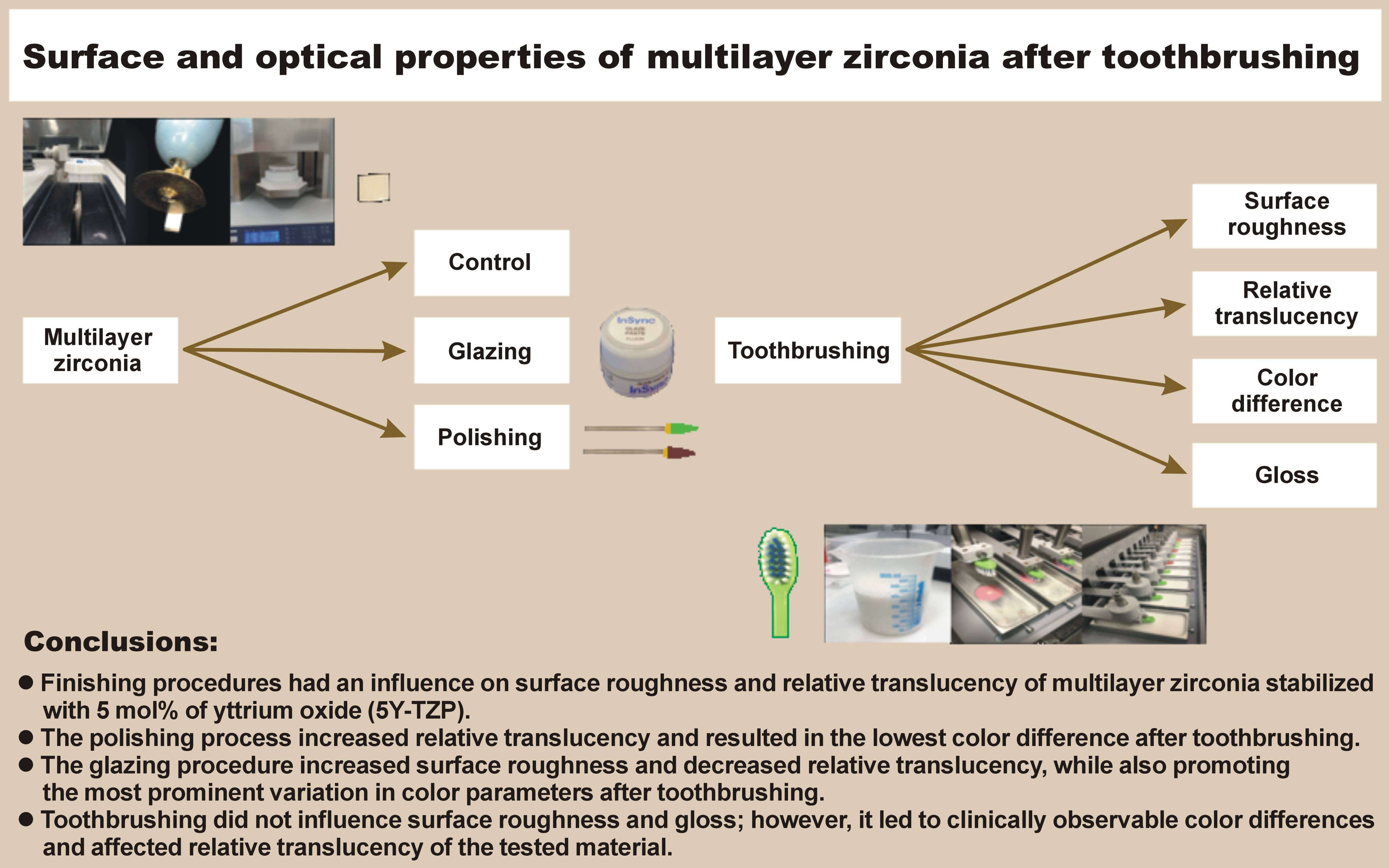
Objectives. The aim of this in vitro study was to evaluate the effect of finishing procedures on the surface roughness (SR) and relative translucency (RT), as well as the effect of toothbrushing on SR, RT, color difference (ΔE00), and gloss (Δgloss) of multilayer zirconia stabilized with 5 mol% of yttrium oxide (5Y-TZP) following polishing or glazing.
Material and methods. Thirty specimens were fabricated from the cervical layer of pre-sintered blocks of 5Y-TZP. The specimens were divided into 3 groups (n = 10/group): control (C); polishing (P); and glazing (G). The surface roughness was evaluated with a confocal laser microscope, and the RT, ΔE00 and Δgloss were assessed with the use of a spectrophotometer. A total of 50,000 cycles of toothbrushing (2 Hz, 2.5 N) were performed using a dentifrice slurry. The linear mixed-effects model and Bonferroni test (α = 0.05) were employed to analyze SR and RT. The color change and Δgloss were assessed using the one-way analysis of variance (ANOVA) and Tukey’s post hoc test (α = 0.05).
Results. The finishing procedures had an influence on SR and RT. The polishing process did not affect SR and increased RT, while the glazing process resulted in an increase in SR and a decrease in RT in the multilayer 5Y-TZP. The impact of toothbrushing on SR was not significant (p = 0.052). However, decreased RT was observed in the P group (p < 0.05), while an increase in RT was noted in the G group (p < 0.001). Additionally, the G group presented the highest mean values for ΔE00, as well as for the difference in lightness (ΔL*), the red/green axis (Δa*) and the yellow/blue axis (Δb*). No statistically significant differences were observed among the groups for Δgloss (p = 0.646).
Conclusions. The polishing process increased RT and resulted in the lowest ΔE00 values after toothbrushing. In contrast, the glazing procedure increased SR and decreased RT, while also promoting the most prominent variation in color parameters after toothbrushing. Toothbrushing with a conventional dentifrice did not influence SR and gloss; however, it led to clinically observable color variations and affected RT of the multilayer 5Y-TZP polycrystals.
Keywords: toothbrushing, color, dental polishing, dental aesthetics, yttria-stabilized tetragonal zirconia
Introduction
Dental zirconia has been increasingly used as a prosthetic restorative material in computer-aided design (CAD) and computer-aided manufacturing (CAD/CAM). The material is widely utilized for tooth- and implant-supported restorations,1 and can be implemented in 2 differ-ent forms: porcelain-veneered zirconia; and monolithic zirconia.2, 3, 4 Failures such as chipping and porcelain fracture have been reported in porcelain-veneered zirconia restorations.2, 3, 4, 5 Monolithic zirconia restorations, which are anatomically contoured restorations fabricated by CAD/CAM, have been proposed to prevent veneer chipping or porcelain fracture, and provide excellent strength with minimal tooth reduction.3, 6, 7, 8 The quality of zirconia-based restorations also depends on the bonding effectiveness of the zirconia surface and the adhesive system. Some authors have reported that bonding effectiveness can be enhanced through the use of single-component universal adhesive9 and laser.10
Zirconia has been marketed with different yttria content. Zirconia partially stabilized with 3 mol% of yttrium oxide (3Y-TZP) exhibits high opacity and is intended for manufacturing frameworks for porcelain-veneered zirconia restorations. Zirconia partially stabilized with 4 mol% of yttrium oxide (4Y-TZP) presents an increase in the yttria content, and, in consequence, increased amount of cubic phase, grain size and translucency. Zirconia stabilized with 5 mol% of yttrium oxide (5Y-TZP) exhibits the highest yttria content, with a maximum of 53% of the cubic phase, which is isotropic. In addition, cubic crystals are larger than tetragonal crystals, which has been shown to reduce the number of grain boundaries that are the source of light scattering. This decrease leads to enhanced translucency in anterior monolithic zirconia restorations.3, 4 In addition, the color of the prosthetic restoration is an important aspect that significantly affects the success of the treatment.11 Monolithic zirconia exhibits a whitish shade; however, matching the aesthetics of natural teeth remains a challenge.12, 13, 14 To address this need, a novel multilayer monolithic zirconia has been developed.
Multilayer zirconia can have a polychromic or hybrid composition. Polychromic multilayer zirconia presents a color gradient by adding different pigments in the cervical and incisal layers of the 5Y-TZP block, while hybrid multilayer zirconia comprises different generations of zirconia in the dentine (3Y-TZP or 4Y-TZP) and enamel (5Y-TZP) layers of the hybrid block.8, 15 A previous study investigated the microstructure, as well as the physical and mechanical properties of polychromic multilayer zirconia and concluded that this material is suitable for fixed prosthetic restorations.16 Monolithic restorations manufactured with the use of polychromic multilayer 5Y-TZP require finishing procedures to improve aesthetics,17 ensure color stability,18 reduce surface roughness (SR),19 minimize biofilm formation,20 and mitigate antagonist wear.21 Although manufacturers recommend both polishing and glazing for monolithic zirconia restorations, there is no well-established method for finishing procedures, and their impact on color difference remains unclear.17, 22 It has been established that 4Y-TZP presents a less enduring glazing layer against toothbrushing compared to reinforced glass ceramics.23
Toothbrushing with a dentifrice is an important component of oral hygiene. However, this process can result in a superficial abrasion and cause color differences in restorative materials.24, 25, 26, 27 Color differences must be evaluated using perceptibility and acceptability thresholds as quality control tools to predict the clinical performance of these restorative materials.28, 29 A number of studies have investigated the effects of toothbrushing on the color difference and SR of 3Y-TZP25, 26, 27, 30, 31 and 5Y-TZP.27, 32 However, the studies that evaluated 5Y-TZP used extrinsic staining, and there is no data concerning the effects of toothbrushing on multilayer 5Y-TZP.
Therefore, the purpose of the present study was to evaluate the effect of finishing procedures on SR and relative translucency (RT), as well as the influence of toothbrushing on SR, RT, color difference (ΔE00), and gloss (Δgloss) of multilayer 5Y-TZP following polishing or glazing. The research hypotheses were as follows: (1) the finishing procedures (polishing or glazing) would have an effect on SR and RT of multilayer 5Y-TZP; and (2) toothbrushing would have an impact on SR, RT, ΔE00, and Δgloss of polished and glazed multilayer 5Y-TZP.
Material and methods
Thirty specimens were fabricated from the cervical layer of 5Y-TZP (ceramill® zolid fx multilayer, LOT 1708000; Amann Girrbach AG, Koblach, Austria) (Table 1). Pre-sintered blocks of 5Y-TZP were cut with a diamond disk (Diamond Wafering Blade; Allied High Tech Products, Inc., Cerritos, USA) in a high precision cutter (IsoMet 1000 Precision Cutter; Buehler, Lake Bluff, USA) under water cooling. The specimens were manually finished with 25-μm grit sandpaper (211Q; 3M ESPE, St. Paul, USA) (pre-sintered specimens’ dimensions: 6.0 mm × 6.0 mm × 1.8 mm), and then sintered in a furnace (inFire HTC Speed; Dentsply Sirona, Charlotte, USA) with a maximum temperature of 1,450°C according to the manufacturer’s instructions (post-sintered specimens’ dimensions: 5.0 mm × 5.0 mm × 1.5 mm).
The sample size was calculated based on the results of a previous study.27 Specimens were divided into 3 groups (n = 10/group) according to the finishing procedure used: control (C); polishing (P); and glazing (G). The finishing procedures were carried out by a single trained operator (LF). Polishing was performed using a medium (W16DC Diacera, LOT 447317; EVE Ernst Vetter GmbH, Keltern, Germany) and fine (W16DCmf Diacera, LOT 446560; EVE Ernst Vetter GmbH) diamond polisher in a slow-speed dental handpiece (Micromotor; Dabi Atlante, Ribeirão Preto, Brazil) operating at 10,000 rpm for 30 s.19, 33, 34 The diamond polisher was positioned on the device for standardization35, 36, 37 and replaced after polishing 5 specimens.31, 36 Glazing was performed by applying a single layer of glaze paste (InSync® Glaze Paste, LOT 172108; Jensen Dental, North Haven, USA) with a brush, followed by firing in a furnace (sinter press alumini; EDG, São Carlos, Brazil) according to the manufacturer’s instructions. The thickness of the specimen was measured with a digital caliper (Absolute Digital Pachymeter; Mitutoyo Corporation, Kawasaki, Japan) before and after glazing to standardize the thickness of the glaze layer (100 µm).27, 36
A confocal laser scanning microscope (CLSM) (LEXT OLS4000; Olympus Corporation, Tokyo, Japan) was used to evaluate the topography and SR before and after 50,000 toothbrushing cycles. A representative image of the surface topography was selected based on the repetitive pattern identified for each group. The surface roughness values (μm) were obtained by analyzing the entire scanned surface using the software dedicated for the CLSM (LEXT OLS4000; Olympus Corporation).
The color difference, RT and gloss were analyzed using a calibrated spectrophotometer (Delta Vista 2.0; Delta Color, São Leopoldo, Brazil) at baseline and after 50,000 toothbrushing cycles. The International Commission on Illumination (CIE) L*, a* and b* color coordinates of the specimens were evaluated. The CIEDE2000 color difference (ΔE00) was calculated based on the previously described formula.38, 39 The relative translucency was obtained by computing the lightness (L*), red/green axis (a*) and yellow/blue axis (b*) values against the white (W) and black (B) backgrounds by using the following formula (Equation 1):
The difference in gloss was calculated using the CIEDE2000 color system based on the following formula (Equation 2):
where:
gloss1* – baseline gloss value;
gloss2* – gloss value measured after toothbrushing.
The specimens were brushed using a linear brushing machine (P200 brushing machine; Biopdi, São Carlos, Brazil) equipped with soft bristle toothbrush heads (J&J REACH Eco; Johnson & Johnson, New Brunswick, USA) using a conventional dentifrice (Colgate Maximum Caries Protection; Colgate-Palmolive, New York, USA) slurry (Table 1). Dentifrice slurries were prepared by mixing distilled water (mL) with a dentifrice (g)25, 27, 30 at a proportion of 2:1 in a vacuum mixer for 2 min. The machine was set at a rate of 180 toothbrushing cycles per minute,25 with a vertical load of 2.5 N23, 25, 32 until 50,000 toothbrushing cycles were completed,27, 32 which simulated 5 years of toothbrushing.23, 27
Statistical analysis
All statistical analyses were performed using the IBM SPSS Statistics for Windows software, v. 20.0 (IBM Corp., Armonk, USA). The Shapiro–Wilk test evaluated the data for normality. Given that the data presented normal distribution, the results of SR and RT were analyzed using repeated measures analysis of variance (ANOVA) and Bonferroni post hoc test (α = 0.05). The data was compared between groups (baseline values) and within groups (before and after toothbrushing) on the same specimens. The results for ΔE00 and Δgloss were analyzed using the one-way ANOVA and Tukey’s post hoc test (α = 0.05).
Results
The finishing procedures had an influence on SR and RT. The recorded baseline mean values indicate that polishing did not influence SR and increased RT, while glazing resulted in an increase in SR and a decrease in RT of multilayer 5Y-TZP (Table 2). The surface roughness of the C and P groups was comparable (p < 0.05). The glazing group presented higher SR compared to the C and P groups (p < 0.001 for both). With respect to RT, the C group exhibited lower mean values than the P group (p < 0.05) and higher results in comparison to the G group (p < 0.001).
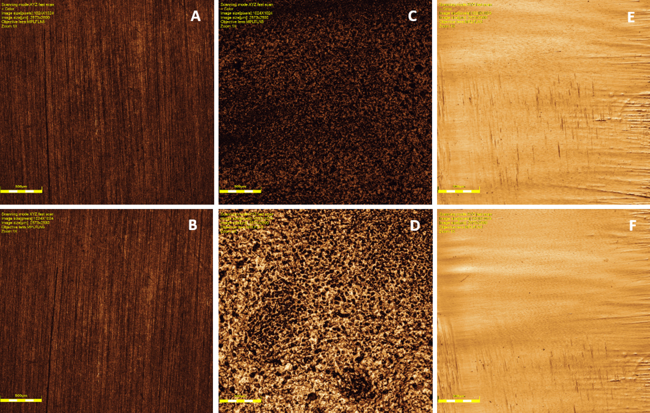
Figure 1 displays representative images of surface topography obtained by means of a laser confocal microscope. At baseline, the G group demonstrated the most irregular surface, while the P group exhibited the most regular one. Toothbrushing had no discernible effect on the surfaces of the C and G groups. However, a reduction in scratches was observed on the surface of the P group.
The impact of toothbrushing on SR was not significant (p = 0.052). However, it resulted in a decrease in RT of the P group (p < 0.05) and an increase in RT of the G group (p < 0.001) (Table 2).
The mean (M) and standard deviation (SD) values of the ΔE00, ΔL*, Δa*, Δb*, and Δgloss for all groups are presented in Table 3. The G group showed the highest mean ΔE00 value. The positive values of ΔL* indicate that toothbrushing increased lightness for all groups. The Δa* and Δb* presented negative values for the C and P groups, and positive values for the G group.
Statistically significant differences were noted among the groups for ΔE00, ΔL*, Δa*, and Δb* values (p < 0.05), and there was no difference for the Δgloss parameter (p = 0.646). The G group presented the highest mean values of ΔE00, ΔL*, Δa*, and Δb*. The C and P groups exhibited similar mean values of ΔE00 (p = 0.064), Δa* (p = 0.337) and Δb* (p = 0.344). Intermediate mean values of ΔL* were noted in the C group, while the ΔL* values in the P group were the lowest (Table 4).
Discussion
The first research hypothesis was accepted because different finishing procedures influenced the baseline SR and RT values of multilayer 5Y-TZP. Glazing resulted in the most irregular surface, high mean values of SR and the lowest RT, while polishing promoted a regular surface and the highest RT.
The second research hypothesis was partially accepted because toothbrushing influenced ΔE00 and RT but did not have an impact on SR and gloss of polished and glazed multilayer 5Y-TZP. In order to ensure the long-term clinical success of aesthetic restorations, it is essential that the restorative materials present color stability and reliable color matching with natural dentition. Color differences can be evaluated with a spectrophotometer and calculated using appropriate mathematical formulas, with values for clinically acceptable color differences being reported. A previous study suggested that the CIEDE2000 color system more accurately represents the human perception of color difference compared to the CIE L*a*b* color space,28 which justifies the use of the CIEDE2000 in this study for the evaluation of visual tolerances. For the CIEDE2000, the 50:50% perceptibility threshold in dentistry was determined to be ΔE00 = 0.8 units, whereas the 50:50% acceptability threshold was found to be ΔE00 = 1.8 units.28, 29 In this study, the observed mean values for color differences after toothbrushing in all groups exceeded the acceptability thresholds. The mean values of ΔE00 for the P, C and G groups can be interpreted as moderately, clearly and extremely unacceptable, respectively.28 The high values for ΔE00 can be attributed to the considerable increase in lightness, indicating that the multilayer 5Y-TZP became luminous after toothbrushing, regardless of the finishing procedure used. Similarly, some authors have reported that monolithic zirconia presented a significant color difference after toothbrushing, with this difference being more pronounced than that observed for glass ceramic.25, 27 Eldwakhly et al. investigated the color differences of dental ceramic specimens subjected to different staining solutions and found that glass ceramic presented the lowest color difference when compared to monolithic zirconia, resin nanoceramic and hybrid ceramic, while monolithic zirconia demonstrated the most substantial color variation.14
It has been reported that 5Y-TZP and 3Y-TZP exhibited different behaviors after toothbrushing, indicating that the chemical composition and crystallographic phase content may influence the color difference of monolithic zirconia.27 In this study, the multilayer 5Y-TZP presented high mean values of color difference, suggesting that the percentage of cubic phase content may have influenced this outcome.
Regarding finishing procedures, Lee et al. investigated the effect of polishing and glazing on intrinsically colored 3Y-TZP that underwent toothbrushing and found that glazed 3Y-TZP presented lower values of color difference after toothbrushing than polished 3Y-TZP.26 In contrast, this study observed that the polished multilayer 5Y-TZP demonstrated lower values of color difference than the glazed multilayer 5Y-TZP. These divergent results can be explained by the method used to color the monolithic zirconia. In the case of intrinsically colored 3Y-TZP, the glazing layer can play a protective role, promoting color maintenance.26 Ataol et al. evaluated the effect of substructure thickness and finishing procedure (polishing or glazing) on the color difference before and after cementation in 3Y-TZP, and found a correlation between substructure thickness and color difference.13 For 0.04-mm substructure thickness, the polished group presented higher mean values of color difference than the glazed group. However, for a substructure thickness of 0.08 mm, the glazed group demonstrated higher mean values than the polished group. In the present study, specimens with the same thickness were used for all groups, and the glazed group presented the highest mean values of color difference. The thickness of the specimens (1.5 mm) is clinically representative because it is similar to the thickness of the occlusal surface of inlays, onlays and crowns recommended by the manufacturer.
There is no consensus regarding the correlation between color differences and SR. Some authors affirmed that color differences can be affected by SR,19 while others argued that there was no correlation between the two.25 In this study, the highest color difference was observed in the glazed group, suggesting that rougher surfaces are related to high mean values of color differences. In addition, the rough surface can lead to plaque accumulation, dental caries, gingival inflammation, and antagonist wear,19, 21 resulting in a decrease in the clinical aesthetic outcome of the prosthesis.17 Sehovic et al. reported that toothbrushing with a conventional dentifrice can lead to an increase in SR of extrinsically stained 3Y-TZP.31 Lee et al. found that extrinsically stained 5Y-TZP showed a decrease in SR after 50,000 toothbrushing cycles, while this study found no differences in SR at baseline and after toothbrushing for glazed and polished multilayer 5Y-TZP for the same number of toothbrushing cycles.32 However, comparisons among these studies are limited because of the different protocols for finishing procedures and toothbrushing, such as the type of toothbrush, machines and load applied.30
Moreover, SR has been demonstrated to affect light reflection.40 After toothbrushing, the polished and glazed groups exhibited different behaviors for RT, but no differences were found in SR. The relative translucency increased in the G group after toothbrushing, while a decrease was observed in the P group. These outcomes may be associated with alterations in the grain size of 5Y-TZP. The grain size had no influence on the SR of 3Y-TZP; however, it could have an influence on RT.41 In addition, Lee et al. investigated the effects of the thickness of extrinsic stain and glazed layers on RT of 5Y-TZP using the CIEDE2000, and concluded that the thickness of extrinsic stain affected RT, but these phenomena were not observed in the glazed group.42
Heintze et al. evaluated the changes in gloss and SR after toothbrushing of restorative materials and found a strong correlation between surface gloss and roughness.43 This study demonstrated that the use of finishing procedures (polishing or glazing) did not influence surface gloss and roughness after toothbrushing using a conventional dentifrice. In contrast, other studies have reported that toothbrushing can lead to a decrease in gloss, depending on the type of dentifrice used.26, 27
The present study demonstrated that finishing procedures influenced SR and RT, and toothbrushing using a conventional dentifrice did not influence SR and gloss, but affected RT and color of polished and glazed multilayer 5Y-TZP. Considering that a finishing procedure should be chosen for monolithic zirconia, the findings of this study suggest that polishing results in a smoother surface and lower mean values of color difference compared to glazing.
Limitations
The limitations of this in vitro study included the fact that toothbrushing cannot simulate the dynamic oral environment, such as pH and masticatory forces. The toothbrushing environment lacked natural compounds found in saliva or chemical insults associated with the intake of meals and beverages. Additionally, dentifrices with different compositions were not investigated. Direct comparisons with other studies are precluded by the presence of different toothbrushing protocols. Our results were limited to polychromic multilayer 5Y-TZP. However, the observed outcomes are relevant because the surface of restorative materials is subjected to toothbrushing with a dentifrice, and the effects of this procedure need to be known.
Further research is necessary to investigate different types of dentifrice, different brands of polychromic multilayer 5Y-TZP and hybrid multilayer zirconia. Additionally, the effect of finishing procedures and toothbrushing on the microstructure and grain size of multilayer zirconia should be investigated. Research efforts should prioritize the development of more stable zirconia to avoid color and translucency variations, considering that these changes have an influence on the success and longevity of dental prosthesis treatment.
Conclusions
Based on the results and within the limitations of this in vitro study, it can be concluded that the application of glazing increased SR and decreased RT, while polishing did not influence SR but increased RT of multilayer 5Y-TZP. The use of a conventional dentifrice during toothbrushing did not influence SR and gloss; however, it led to clinically observable color differences and affected RT of the tested material.
Ethics approval and consent to participate
Not applicable.
Data availability
The datasets generated and/or analyzed during the current study are available from the corresponding author on reasonable request.
Consent for publication
Not applicable.
Use of AI and AI-assisted technologies
Not applicable.