Dental and Medical Problems

Dent Med Probl
Impact Factor (IF 2024) – 3.9
Journal Citation Indicator (JCI 2024) - 1.36
Scopus CiteScore (2024) – 5.0
Index Copernicus Value (ICV 2023) – 181.00
MNiSW – 70 pts
ISSN 1644-387X (print)
ISSN 2300-9020 (online)
Periodicity – bimonthly


Download original text (EN)

Dental and Medical Problems

2023, vol. 60, nr 3, July-September, p. 527–535

doi: 10.17219/dmp/140964

Publication type: review

Language: English

License: Creative Commons Attribution 3.0 Unported (CC BY 3.0)

Download citation:

  • BIBTEX (JabRef, Mendeley)
  • RIS (Papers, Reference Manager, RefWorks, Zotero)

Cite as:


Yazarloo S, Arab S, Mirhashemi AH, Gholamrezayi E. Systematic review of preventive and treatment measures regarding orthodontically induced white spot lesions. Dent Med Probl. 2023;60(3):527–535. doi:10.17219/dmp/140964

Systematic review of preventive and treatment measures regarding orthodontically induced white spot lesions

Samira Yazarloo1,A,B, Sepideh Arab2,D,E,F, Amir Hossein Mirhashemi2,C,E, Elahe Gholamrezayi2,B,D

1 Department of Orthodontics, Shahid Sadoughi University of Medical Sciences, Yazd, Iran

2 Department of Orthodontics, School of Dentistry, Tehran University of Medical Sciences, Iran

Abstract

White spot lesions (WSLs) are one of the most common adverse effects following comprehensive fixed orthodontic treatment. The purpose of this review was to evaluate recent studies addressing the prevention and treatment of these lesions. Electronic databases were searched for English-written studies published between 2015 and October 2020 involving randomized clinical trials aiming at prevention or treatment of orthodontically induced WSLs using the following keywords in their title or abstracts: randomized clinical trial OR randomized controlled trial AND white spot OR caries OR demineralization OR decalcification OR remineralization. From the 23 papers which met the inclusion criteria, 11 were on preventive methods, while 12 addressed treatment protocols. However, most of the reviewed studies had a high risk of bias. The results of this review strongly support the importance of oral hygiene observation in preventing WSLs. Sodium fluoride varnish 5% was confirmed to be effective in the treatment of these lesions, as well as in the prevention of WSLs in patients with suboptimal oral hygiene. In addition, immediate CO2 laser irradiation after bonding can effectively prohibit formation of WSLs during orthodontic treatment. The literature also illustrates a promising masking effect of resin infiltration for the treatment of WSLs. However, little scientific evidence supports the effectiveness of Casein phosphopeptide-amorphous calcium phosphate (CPP-ACP) against WSLs, although more clinical trials with long-term follow-up are needed. Oral hygiene maintenance is crucial in the prevention of WSLs, and 5% sodium fluoride varnish and CO2 laser irradiation are recommended in patients with compromised oral hygiene. In the case of WSL formation, fluoride varnish and resin infiltration are effective treatment modalities.

Keywords: dental caries, tooth remineralization, fixed orthodontic appliances, randomized controlled trial

Introduction

A common and unpleasant consequence of fixed orthodontic treatments is the development of white spot lesions (WSLs). These are characterized by white opaque lesions that are formed as a result of increased enamel subsurface porosities, which are early and reversible caries.1, 2, 3 While regular caries take at least 6 months to develop, WSLs may become visible within a month following bonding of attachments.4

Since one of the main goals of orthodontic treatment is to improve smile esthetics, WSLs can negatively affect treatment outcomes and lead to patient dissatisfaction,5 and they have a prevalence of 4.9 to 84%. This wide range of prevalence could be attributed to different study designs and the variety of methods used for detection and quantification of demineralized areas.6 Although almost every tooth can be affected, most of the studies report higher involvement rates in the lateral incisors.7, 8, 9

Improvement of WSLs appearance mostly occurs in the first 6-24 months after deboning. This is due to three major factors: oral hygiene improvement and reduction in Streptococcus mutans and Lactobacillus spp to baseline level, remineralization using fluoride-containing dentifrices and mouth rinses, and abrasion of lesions due to brushing.10, 11

Several interventions have been introduced for WSL management. Some are based on remineralization using high concentration fluoride components such as varnish, mouth rinse, and casein phosphopeptide-amorphous calcium phosphate (CPP-ACP) (MI paste/ MI paste plus), while others focus on improvement of appearance by other methods like bleaching, micro-abrasion, and resin infiltration.12, 13

The aim of this literature review was to investigate the latest randomized clinical trials in order to clarify evidenced-based measures for prevention and treatment of WSLs during and after fixed orthodontic treatment.

Methods

Protocol and registration

The current systematic review followed the PRISMA guidelines14 and the Cochrane Handbook for the Systematic Review of Interventions (version 5.1.0).15

Eligibility criteria

According to the PICOS format (Table 1), we included randomized controlled trials on human patients comparing various preventive/therapeutic measures addressing orthodontically induced WSLs to a control/placebo group.

The eligibility criteria are summarized below.

Inclusion criteria:

– randomized clinical trial studies;

– treatment by conventional fixed orthodontic appliances;

– at least one post-orthodontic WSL (trials concerning WSL treatment)

– studies investigating different preventive and therapeutic strategies except veneers; and

– papers published in English between 2015 and October 2020.

Exclusion criteria:

– other designs of studies;

– animal studies and in vitro studies; and

– WSLs not stated to be the consequence of orthodontic treatment

Information sources and search strategy

A comprehensive electronic search was performed in PubMed, Scopus, and Embase databases from 2015 to October 2020 using the following keywords in their title or abstracts: randomized clinical trial OR randomized controlled trial AND white spot OR caries OR demineralization OR decalcification OR remineralization (Table 2). We manually added suitable articles for inclusion, and the reference lists were screened for new trials.

Study selection and data extraction

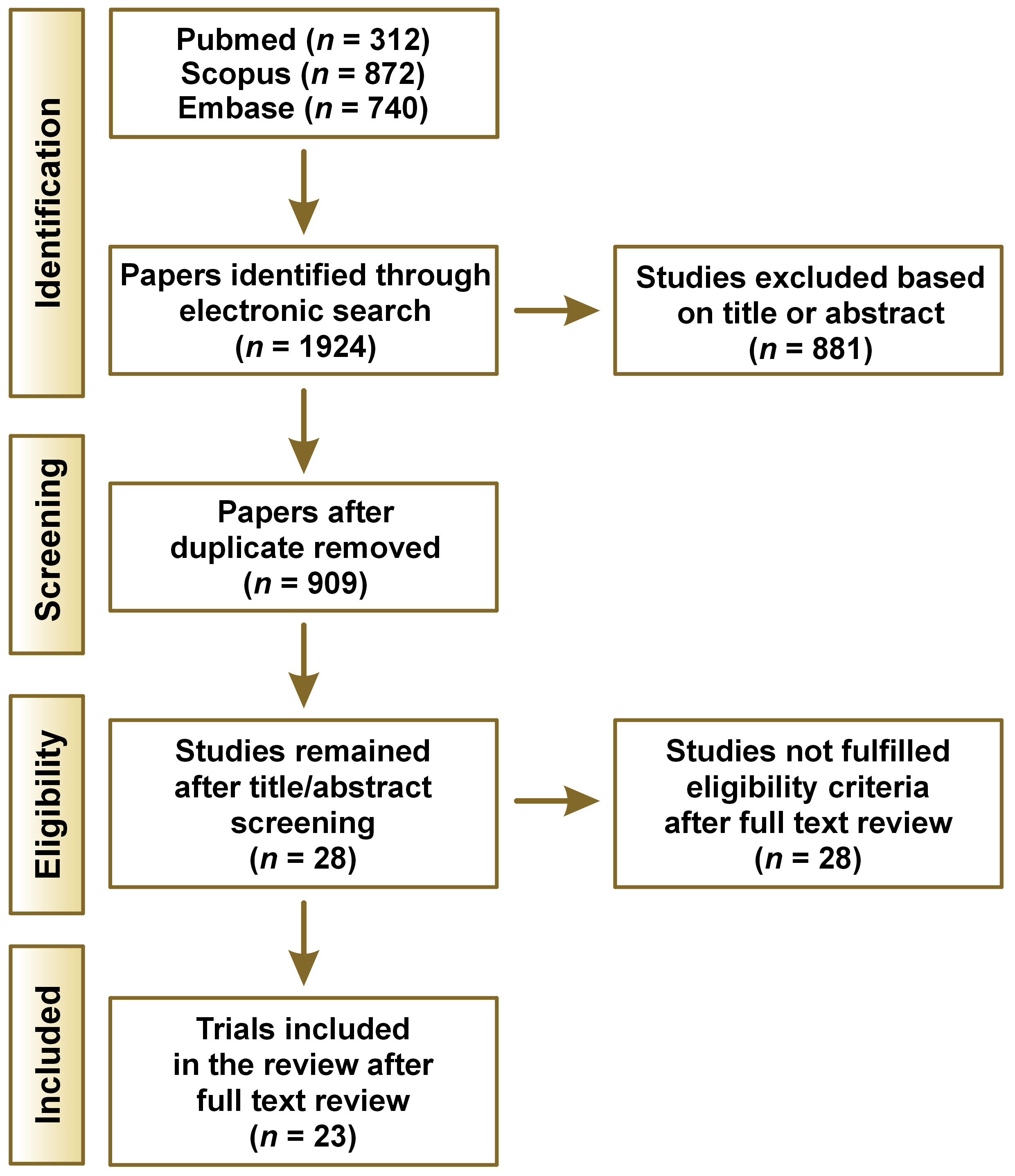
A total of 1924 papers were identified through the electronic search. After duplicate papers were removed by EndNote X9, 909 papers remained. Afterwards, two investigators independently evaluated the titles and abstracts considering the inclusion and exclusion criteria. Inconsistencies between reviewers were excluded from the study unless a consensus was reached. Full texts of 28 relevant papers were selected and reviewed thoroughly, and 23 articles met the final criteria for entering this review (Figure 1). We extracted the following data from the included studies to facilitate comparison among the articles: number of participants, type of intervention, follow-up time, diagnostic method used, and conclusions.

Assessment of the risk of bias in the included studies

Two review authors independently assessed the risk of bias for the included trials using Cochrane’s risk of bias tool.15 The study was judged as low risk if bias of all the domains were low. If at least one domain was considered high risk, the study was judged as high risk. A moderate risk of bias was considered if at least one domain was judged as unclear.

Results

Twenty-three papers met the qualifications for this systematic review. The literature involved both prevention and treatment of WSLs during and after orthodontic treatment.

Eleven papers addressed preventive strategies. One study was on bonding agents (fluoride-containing adhesive resin),16 six on fluoride/ CPP-ACP containing products (varnish, toothpaste, mouth rinse, MI Paste Plus , etc.),17, 18, 19, 20, 21, 22, 23 one study on photodynamic therapy,24 one on special sealant,25 and two studies were on CO2 laser.26, 27

Twelve papers considered therapeutic strategies. Of these, seven studies were on remineralizing agents,28, 29, 30, 31, 32, 33, 34 two studies were on resin infiltration,35, 36 one study evaluated the effectiveness of resin infiltration and microabrasion,37 one study compared fluoride varnish and chlorhexidine mouthwash, and one study compared resin infiltration with varnish.38 Table 3 demonstrates the general characteristics of the twenty-three included papers.

Risk of bias assessment

The details of the quality assessment of twenty-three eligible studies included in this systematic review were extracted and assessment of the risk of bias for included studies was performed. Two studies were assessed to be low, two studies were considered to be moderate, and nineteen studies were classified as high risk of bias (Table 4).

Discussion

WSL development is a common adverse effect following fixed orthodontic treatment which compromises the esthetic outcomes of treatment. Therefore, prevention and management of WSLs is extremely important for clinicians. In this review, we assessed recently published randomized clinical trials concerning both prevention and treatment of post-orthodontic WSLs.

Despite the breadth of literature concerning this issue, there is still no consensus on how to prevent or treat post-orthodontic WSLs. However, it is always easier and more effective to prevent rather than treat.

The current literature contains invaluable reviews concerning white spot lesions.39, 40, 41, 42, 43 However, some of these have focused on a single remineralizing agent,39, 42, 43 and the others were limited to assessing remineralizing agents without addressing contemporary approaches such as laser irradiation,40, 41, 43 and some were not focused on orthodontically induced lesions.39, 40 Another advantage of the current study was the evaluation of both preventive and treatment approaches together. In addition, the current study summarized further information such as location of the lesions and age group of the participants (Table 3). This shows that most of the included studies evaluated the labial surfaces of the teeth with brackets, which is sensible due to the prevalence and mechanism of white spot lesions. However, the studies were limited to patients under 30 years old, which reveals the need for studies on prevention and treatment of lesions in older patients. In the current study, we tried to overcome the shortages with a more comprehensive review.

The diversity in the results from clinical trials can be due to variable methods and study designs, a wide range of products claiming effectiveness on WSL treatment, various methods for the detection and quantification of WSLs, and different procedures for the application of active agents. Visual detection indices including International Caries Detection and Assessment System (ICDAS) and EDI, spectrophotometer, DIAGNOdent, and QLF are the most common methods used for detection and quantification of WSLs. Using the naked eye is not a reliable indicator, whereas magnifying loupes accompanying air drying of tooth surface is an effective method for identifying WSLs of smooth surfaces. However, there is no significant difference between DIAGNOdent and loupes.44 Since all studies using visual indices for detecting or assessing improvement of WSLs has depended on the naked eye only and did not incorporate magnifying loupes, minor improvement of lesions might have been ignored which can directly influence the studies’ results.

Prevention

Results of this review emphasize the pivotal role of routine oral hygiene maintenance in prevention of WSLs. Regular brushing with two different fluoride dentifrices (Clinpro 5000 and Clinpro Tooth Creme) twice daily for two minutes was effective in hindering WSLs, with results comparable to those of MI Paste Plus.20 The importance of routine oral hygiene procedures is also noted by Rechmann et al. who demonstrated that the combination of fluoride tooth paste and fluoride varnish and MI Paste Plus together does not provide any preventive advantage over the routine use of fluoride tooth paste and mouth rinse. In addition, it has been shown that using 5% sodium fluoride varnish is effective against WSL formation only in patients with suboptimal oral hygiene, and it is not useful in those with perfect oral hygiene.19 Similarly, a single dose of elmex® fluid (10,000 ppm) and Fluor Protector S (7700 ppm) had no benefit over the routine sufficient dental hygiene approach, and application of 1.5% ammonium fluoride every six weeks only reduced the number of severe lesions (which are more likely to be sighted in patients with poor oral hygiene).13, 17

Several interventions such as photodynamic therapy, using ultrasonic scaler and the application of sealants have been tested for their capability against WSLs. However, none of them were successful in the prevention of WSLs.24, 25 Resin Modified Glass Ionomer cement (RMGIC) has shown promising results in preventing WSL development during orthodontic treatment.22 However, one split mouth RCT revealed no preventive effect of fluoride-releasing resin composite against demineralization and development of WSLs adjacent to brackets.16

There is ample evidence that laser irradiation makes enamel more resistant to acid-induced demineralization by altering its structure.45 Various types of lasers have been introduced for this purpose, including CO2, erbium-doped yttrium aluminum garnet (Er: YAG), neodymium-doped yttrium aluminum garnet (Nd: YAG), erbium, chromium: yttrium-scandium-gallium-garnet (Er, Cr: YSGG), diode, and argon lasers. However, studies that assessed laser effectiveness in reducing enamel susceptibility were mainly in vitro and were not included in this review.46 There is supporting data for effectiveness of CO2 laser in the prevention of WSL formation. In 2019, Mahmoudzadeh et al. exposed teeth to CO2 laser (0.4 mw, 10.6 μm, 5 Hz) for 20 s following bracket attachment and uncovered that CO2 laser irradiation not only caused a reduction in WSL incidence, but it also diminished their extent and severity after 6 months.26 Raghis et al. also concluded that CO2 laser irradiation had an inhibitory effect on WSL formation during orthodontic treatment after 2 and 6 months.27 The effectiveness of laser irradiation in preventing enamel demineralization was evaluated in a systematic review in 2018; however, more RCTs are needed to verify the clinical efficacy of different available systems.45 In addition, the cost-effectiveness of using lasers should be assessed in comparison to the more economical and accessible traditional approaches, such as using fluoride varnish.

Treatment

Careful supervision and waiting for at least 3-6 months until natural remineralization occurs is the method of choice in most patients. The optimal use of fluoride-containing toothpastes, which is twice daily without excessive rinsing after brushing, must be carefully monitored during this period.13, 47, 48 However, natural remineralization is insufficient in many cases, and sometimes it doesn’t take place at all. Therefore, in many patients, adjunctive considerations should be considered.

Results of this review revealed that MI Paste Plus does not provide any long term benefits in the treatment of WSLs, while short term application of it has shown controversial results.28, 34 All reviewed clinical trials strongly support using 5% sodium fluoride varnish as an effective treatment for WSLs.29, 30, 35, 36, 37, 38

In 2018, Fernandez et.al concluded that most remineralizing agents were not more effective than conventional oral hygiene protocols, and the only effective product for remineralization of WSLs was 5% sodium fluoride varnish applied professionally once per month for 6 months.49 Although application of fluoride varnish on a monthly basis can effectively lead to reversal of WSLs after debonding, the use of high concentration fluoride immediately after debonding is under question, perhaps because surface hyper-mineralization may arrest remineralization and increase the risk of permanent brown discoloration.2, 50

Despite the positive results of fluoride varnish, the effectiveness of using 1.25% fluoride gel for treatment of WSLs is not supported by the literature.32, 33 Application of ACP-CPP alone has not shown significant improvement of WSLs, although it is effective when combined with daily use of fluoride dentifrices.29, 31

Similar to fluoride varnish, resin infiltration has also shown successful results for the treatment of WSLs. Comparison of these two methods demonstrated that resin infiltration shows greater improvement immediately after intervention, while fluoride varnish displays significantly better results over time.38 One split mouth study that compared resin infiltration with microabrasion illustrated esthetic improvement of WSLs with both techniques, but resin infiltration showed better enhancement at 12 months.37

None of the included studies compared microabrasion with a control group. Only one paper made a comparison between resin infiltration and microabrasion as discussed above.37 Microabrasion is an invasive method which is capable of masking more severe and long-standing lesions; however, care must be taken, as this is a sensitive method and should be repeated several times.51 No recent studies concerning bleaching of post-orthodontic WSLs fulfilled the inclusion criteria.

Conclusions

Observation of oral hygiene by brushing with fluoride toothpaste is the backbone of prevention of WSLs in patients undergoing orthodontic treatment. However, application of 5% fluoride varnish or using a CO2 laser after bonding are recommended in patients with compromised oral hygiene. If WSLs are formed during fixed orthodontic treatment, fluoride varnish 5% and resin infiltration are effective methods for treatment.

Ethics approval and consent to participate

Not applicable.

Data availability

The datasets generated and/or analyzed during the current study are available from the corresponding author on reasonable request.

Consent for publication

Not applicable.

Tables


Table 1. PICO format of the review

Population

Patients with fixed orthodontic treatment, with post orthodontic white spot lesions

Intervention

Various preventive/therapeutic measures for WSLs (except veneers)

Comparison

No intervention, normal home care

Outcome

Effects on mineral content, appearance, or extent of lesions

Table 2. Search strategy

Pubmed

(((((white spot[Title/Abstract]) OR (decalcification[Title/Abstract])) OR (remineralization[Title/Abstract])) OR (demineralization[Title/Abstract])) OR (caries[Title/Abstract])) AND ((randomized clinical trial[Title/Abstract]) OR (randomized controlled trial[Title/Abstract])) Filters: from 2015 – 2020

Scopus

((TITLE-ABS-KEY (white AND spot) OR TITLE-ABS-KEY (caries) OR TITLE-ABS-KEY (demineralization) OR TITLE-ABS-KEY (remineralization) OR TITLE-ABS-KEY (decalcification))) AND ((TITLE-ABS-KEY (randomized AND clinical AND trial) OR TITLE-ABS-KEY (randomized AND controlled AND trial))) AND (LIMIT-TO (PUBYEAR , 2021) OR LIMIT-TO (PUBYEAR , 2020) OR LIMIT-TO (PUBYEAR , 2019) OR LIMIT-TO (PUBYEAR , 2018) OR LIMIT-TO (PUBYEAR , 2017) OR LIMIT-TO (PUBYEAR , 2016) OR LIMIT-TO (PUBYEAR , 2015)) AND (LIMIT-TO (SUBJAREA , "DENT"))

Embase

'white spot' OR 'caries'/exp OR caries OR 'decalcification'/exp OR decalcification OR 'demineralization'/exp OR demineralization OR 'remineralization'/exp OR remineralization AND 'randomized clinical trial' OR 'randomized controlled trial'/exp OR 'randomized controlled trial' OR rct AND (2015:py OR 2016:py OR 2017:py OR 2018:py OR 2019:py OR 2020:py OR 2021:py) AND ('adverse device effect'/dm OR 'adverse event'/dm OR 'demineralization'/dm OR 'dental caries'/dm OR 'dentin sensitivity'/dm OR 'gingiva bleeding'/dm OR 'gingiva disease'/dm OR 'gingivitis'/dm OR 'malocclusion'/dm OR 'periodontal disease'/dm OR 'periodontitis'/dm OR 'pulpitis'/dm OR 'tooth disease'/dm OR 'tooth pain'/dm OR 'tooth plaque'/dm OR 'tooth pulp disease'/dm) AND 'randomized controlled trial'/de

Table 3. Summary of the included studies

No.

Study

Number of participants

Age

Areas studied/
location of lesions

Caries risk assessment

Type of intervention

Duration

Diagnostic method

Conclusions

Prevention

1

Kumar
et al 2015

40

12–20 years

incisors and canines

-

RMGI varnish
(split mouth)

6 months

DIAGNOdent direct visual inspection

resin-modified glass ionomer cement varnish had an effective role in prevention of WSLs during orthodontic treatment

2

Kirschneck
et al 2016

90

10–17 years

all maxillary and
mandibular dental surfaces with orthodontic brackets

ICDAS index of <2

placebo varnish (30)
elmex® (30) (10000ppm)
Fluor Protector S (30) (7700ppm)

4 weeks 12 weeks 20 weeks

ICDAS

Not any significant difference
in the mean ICDAS in 3 groups /
ICDAS index increased in 3 groups in T3

3

Hammad
et al 2016

50

12–18 years

all maxillary and
mandibular dental surfaces with orthodontic brackets

adequate oral hygiene

SeLECT DefenseTM sealant (25)
Control (25)

12 months

Plaque Index (API)

sealant showed no significant effect as a
solo preventive strategy.

4

Perrini
et al 2016

24

mean age of
14 years 1 month.

Anterior, Middle, Posterior teeth

-

5% Varnish every 3 months (13)
Varnish applied every 6 months (11)
Controls (Split mouth)

3 months
6 months
9 months
12 months

DIAGNOdent

Periodic application of varnish can offer some protection against
WSLs, but not to a statistically significant degree if the patients have excellent oral hygiene

5

Alabdullah
et al 2017

34

13–25 years

all maxillary and
mandibular dental surfaces with orthodontic brackets

adequate oral
hygiene

fluoride-containing adhesive
resin
control
(split mouth)

3 months
6 months
9 months
12 months

photograph
Visual-inspection
DIAGNOdent

Fluoride-containing resin adhesive does not have the desired preventive effect to prevent demineralization and WSL formation

6

Gomez
et al 2018

20

12-18 years

Tooth surfaces with an orthodontic bracket

ICDAS index <1

Photodynamic Therapy (10)
ultrasonic scaler (10)

3 months
6 months
9 months

ICDAS

No significant difference in the mean ICDAS Index between two groups / index slightly increased from T2 follow-up in both groups

7

Rechmann
et al 2018

40 (37 for follow up)

11 years and
Older

facial surfaces of incisors, canines, and first bicuspids

EDI mean: control 37.7
Experimental 42.9
ICDAS mean: Control 21.1
Experimental 21.9

Toothpaste + varnish (every 3 months) + MI paste plus (19)
Toothpaste + mouthrinse (control) (18)

3 months
6 months
12 months

EDI
ICDAS

no statistically significant differences in EDI sum and ICDAS
scores between 2 groups

8

Raghis
et al 2018

26

14 and 25 years

-

good oral
hygiene

10.6 μm CO2 laser irradiation after bonding
Control non-therapeutic light
(split mouth)

1 month
2 month
6 month

clinical and photographic
examinations
digital images DIAGNOdent

10.6 μm CO2 laser has an inhibitory
effect on demineralized lesions formation during orthodontic treatments.

9

Kau
et al 2019

120 (100 for follow up)

12 years and
Older

Facial surfaces of up to 20 teeth evaluated for enamel decalcification.

EDI mean:
Clinpro 5000: 100
Clinpro Cr`eme: 97.7
MI Paste: 99.8

Clinpro 5000 (35)
Clinpro Tooth Crème (32)
MI paste plus (33)

1 month
2 months
3 months
4 months

Enamel Decalcification Index (EDI)

All treatments have a reduction effect on white spot lesions/ Clinpro 5000 showing a marginally better effect
than the two other

10

Mahmoudzadeh
et al 2019

95

12–30 years

Maxillary anterior teeth

-

0.4 mw, 10.6 μm, 5 Hz for 20 s, CO2 laser after bonding (278)
placebo light control (276)

6 months

Visual inspection (WSLs’ incidence)
EDI (WSLs’ extent)
Clinical assessment (WSLs’ severity)

CO2 laser effectively prevent incidence of WSLs, its effectiveness varied depending on the surface region.

11

Sonesson
et al2020

182 (148 completed the trial)

12–18 years

bonded maxillary teeth

-

Varnish Ammonium fluoride 1.5% (75) every 6 week
Placebo varnish (73)

Until the end of treatment

Digital photos

Ammonium fluoride reduces the prevalence of severe lesions (score 3 and 4).

Treatment

12

Eckstein
et al2015

20 (9 for follow up)

13-19 years

Quadrants with noncavitated, postorthodontic WSLs

Resin infiltration
(split-mouth)

6 months
12 months

Spectrophotometer

esthetic improvement of postorthodontic WSL with resin infiltration

13

Singh
et al 2016

41

16-25 years

labial surface of each tooth in both the jaws from central
incisor to the first molar on either side

Control/ Fluoride Toothpaste (14)
Toothpaste + varnish5 % (13)
Toothpaste + CPP-ACP plus(14)

1 month
3 months
6 months

DIAGNOdent

Twice daily application of fluoride varnish or CPP-ACP plus crème along with fluoride toothpaste for 6 months has significant effect on remineralization of WSLs and decreased the severity of WSLs
No additional benefit from using fluoride varnish and CPP-ACP

14

Restrepo
et al 2016

35

13-20 years

Facial surfaces of anterior teeth and premolars

5% NaF varnish (12 patients, 2 apps one-week interval) 2% chlorhexidine gel (12 patients, 2 apps one-week interval)
home care control(11 patients)

1 week
1month
2 months
3 months

DIAGNOdent
Nyvad criteria

F and CHX were capable of controlling the
WSLs adjacent to bracket/ F induced faster remineralization than CHX

15

He
et al 2016

240 (211 for follow up)

12-25 years

All maxillary anterior teeth with a WSL but no cavitary
caries

Fluoride varnish 5%(69)
Fluoride film (Sheer) 5% (70)
control (72)

3 months
6 months

Quantitative light-induced fluorescence

Treatment with either fluoride varnish or film can induce the greatest amount of remineralization in comparison with control group. Fluoride varnish may be slightly more effective than fluoride film.

16

Karabekiroglu
et al 2017

41 (34 for follow up)

14-20

buccal surfaces of the anterior teeth, premolars,
and first molars in the upper and lower jaws

CPP-ACP (16)
Control (18)

36 months

DIAGNOdent
Gorelick index
ICDAS II criteria

Daily application of ACP-CPP was not significantly effective

17

Bock et al
2017

46 (39 for follow up)

>11 years

Four upper front teeth

1.25 per cent fluoride gel (21)
Control (18)

1 week
2 weeks
6 weeks
12 weeks
24 weeks

digital intraoral photographs (WSL dimension)
(WSL luminance)

no significant positive effect of high-dose fluoride on post-orthodontic WSL development (based on a photographical assessment of WSL luminance)
Dimensional WSL quantification showed limited reliability

18

Bock et al
2017

46 (39 for follow up)

>11years

Four upper front teeth

1.25 per cent fluoride gel (21)
Control (18)

1 week
2 weeks
6 weeks
12 weeks
24 weeks

Modified WSL index
Lesion activity assessment (LAA)

No significant positive effect of high-dose fluoride (1.25 percent) on clinical post-orthodontic WSL was seen when compared with the placebo

19

Heravi et al
2018

36

13-23 years

Labial surfaces of six maxillary anterior teeth

MI paste plus (CPP-ACP and F) (12)
Remin Pro (Hydroxyapatite and F) (12)
Control (12)

4 weeks
8 weeks
12 weeks

Fluorescent camera

Both treatments are effective in reducing the area, increasing the mineral content, improving the appearance

20

Beerens et al
2018

51

12-19 years

Buccal surfaces of maxillary and mandibular teeth

MI Paste Plus (25)
Placebo (26)

6 weeks
3 months

6 months
12 months

quantitative light-induced fluorescence (QLF)
lesion changes scored visually

a significant improvement in enamel lesions over time in both groups with no differences between groups

21

Kannan et al
2019

12

14-30 years

Teeth with WSL after removal of fixed orthodontic appliances

Icon® resin infiltration (6)
Clinpro™ XT varnish (6)

immediately
3 months
6 months

Spectrophotometer
DIAGNOdent

Immediately after the intervention, Icon® resin infiltration demonstrated a significantly better improvement than Clinpro™ XT varnish. Conversely, at 3 and 6 months, Clinpro™ XT varnish showed significantly better improvement.

22

Knosel et al
2019

20 (8 for follow up)

12-17 years

Nonrestored, noncavitated postorthodontic WSL after multibracket treatment

Resin infiltration
(split-mouth)

6 months
12 months
+24 months

Spectrophotometer

Suitable for long term esthetic appearance improvement

23

Gu et al
2019

20 (16 available at T 12)

12-19 years

Anterior maxillary or mandibular teeth with WSLs

Resin infiltration (54)
Microabrasion (54)
Control (108)(split-mouth)

1 week
6 months
12 months

Photographs
Spectrophotometer

Improved appearance of WSL with resin infiltration and microabrasion/ higher effect of resin infiltration at 12months

Table 4. Summary of the risk of bias for RCT studies according to thethe Cochrane Collaboration tool for assessing risk of bias

No.

Study

Design

Random sequence generation

Allocation concealment

Blinding of outcome assessment

Incomplete outcome data

Selective reporting

Risk
of bias

1

Heravi et al

RCT

Low

Low

Low

Low

Unclear

Moderate

2

Karabekiroglu et al

RCT

Low

High

Low

High

Unclear

High

3

Singh et al

RCT

Low

Low

Low

High

Low

High

4

Gu et al

RCT

Low

Low

Low

High

Low

High

5

Knosel et al

RCT

Low

High

High

High

Low

High

6

Eckstein et al

RCT

Low

High

High

High

Low

High

7

He et al

RCT

Low

Low

Low

High

Low

High

8

Bock et al

RCT

Low

Low

Low

High

Low

High

9

Beerens et al

RCT

Low

Low

Low

High

High

High

10

Kannan et al

RCT

Low

High

Unclear

High

Low

High

11

Bock et al

RCT

Low

Low

Low

High

Low

High

12

Restrepo et al

RCT

Low

Low

Low

High

High

High

13

Kumar et al

RCT

Low

Unclear

Low

High

High

High

14

Kirschneck et al

RCT

Low

Low

Low

Low

Low

Low

15

Perrini et al

RCT

Low

High

Unclear

Low

Low

High

16

Gomez et al

RCT

Low

Low

Low

High

High

High

17

Kau et al

RCT

Low

Low

Unclear

High

High

High

18

Hammad et al

RCT

Low

Low

Low

High

Unclear

High

19

Rechman et al

RCT

Low

Low

Low

High

Unclear

High

20

Alabdullah et al

RCT

Low

Low

Low

High

Low

High

21

Raghis et al

RCT

Low

Low

Low

Low

Low

Low

22

Mahmoudzadeh et al

RCT

Low

Low

Low

High

Unclear

High

23

Sonesson et al

RCT

Low

Low

Low

Low

Unclear

Moderate

Figures


Fig. 1. Flow diagram of the study

References (51)

  1. Bishara SE, Ostby AW. White spot lesions: Formation, prevention, and treatment. Semin Orthod. 2008;14(3):174–182. doi:10.1053/j.sodo.2008.03.002
  2. Du M, Cheng N, Tai B, Jiang H, Li J, Bian Z. Randomized controlled trial on fluoride varnish application for treatment of white spot lesion after fixed orthodontic treatment. Clin Oral Investig. 2012;16(2):463–468. doi:10.1007/s00784-011-0520-4
  3. Bailey DL, Adams GG, Tsao CE, et al. Regression of post-orthodontic lesions by a remineralizing cream. J Dent Res. 2009;88(12):1148–1153. doi:10.1177/0022034509347168
  4. Øgaard B. White spot lesions during orthodontic treatment: Mechanisms and fluoride preventive aspects. Semin Orthod. 2008;14(3):183–193. doi:10.1053/j.sodo.2008.03.003
  5. Bröchner A, Christensen C, Kristensen B, et al. Treatment of post-orthodontic white spot lesions with casein phosphopeptide-stabilised amorphous calcium phosphate. Clin Oral Investig. 2011;15(3):369–373. doi:10.1007/s00784-010-0401-2
  6. Downer MC, Blinkhorn AS, Holt RD, Wight C, Attwood D. Dental caries experience and defects of dental enamel among 12‐year‐old children in north London, Edinburgh, Glasgow and Dublin. Community Dent Oral Epidemiol. 1994;22(5 Pt 1):283–285. doi:10.1111/j.1600-0528.1994.tb02051.x
  7. Mizrahi E. Surface distribution of enamel opacities following orthodontic treatment. Am J Orthod. 1983;84(4):323–331. doi:10.1016/s0002-9416(83)90348-2
  8. Baeshen HA, Lingström P, Birkhed D. Effect of fluoridated chewing sticks (miswaks) on white spot lesions in postorthodontic patients. Am J Orthod Dentofacial Orthop. 2011;140(3):291–297. doi:10.1016/j.ajodo.2010.04.034
  9. Chapman JA, Roberts WE, Eckert GJ, Kula KS, González-Cabezas C. Risk factors for incidence and severity of white spot lesions during treatment with fixed orthodontic appliances. Am J Orthod Dentofacial Orthop. 2010;138(2):188–194. doi:10.1016/j.ajodo.2008.10.019
  10. Rosenbloom RG, Tinanoff N. Salivary Streptococcus mutans levels in patients before, during, and after orthodontic treatment. Am J Orthod Dentofacial Orthop. 1991;100(1):35–37. doi:10.1016/0889-5406(91)70046-Y
  11. Shungin D, Olsson AI, Persson M. Orthodontic treatment-related white spot lesions: A 14-year prospective quantitative follow-up, including bonding material assessment. Am J Orthod Dentofacial Orthop. 2010;138(2):136.e1–e8. doi:10.1016/j.ajodo.2009.05.020
  12. Welbury RR, Carter NE. The hydrochloric acid-pumice microabrasion technique in the treatment of post-orthodontic decalcification. Br J Orthod. 1993;20(3):181–185. doi:10.1179/bjo.20.3.181
  13. Sonesson M, Bergstrand F, Gizani S, Twetman S. Management of post-orthodontic white spot lesions: An updated systematic review. Eur J Orthod. 2017;39(2):116–121. doi:10.1093/ejo/cjw023
  14. Liberati A, Altman DG, Tetzlaff J, et al. The PRISMA statement for reporting systematic reviews and meta-analyses of studies that evaluate health care interventions: Explanation and elaboration. J Clin Epidemiol. 2009;62(10):e1–e34. doi:10.1016/j.jclinepi.2009.06.006
  15. Higgins JPT, Green S, eds. Cochrane Handbook for Systematic Reviews of Interventions. Version 5.1.0 (updated March 2011). The Cochrane Collaboration, 2011. https://www.handbook.cochrane.org. Accessed February 20, 2021.
  16. Alabdullah MM, Nabawia A, Ajaj MA, Saltaji H. Effect of fluoride-releasing resin composite in white spot lesions prevention: A single-centre, split-mouth, randomized controlled trial. Eur J Orthod. 2017;39(6):634–640. doi:10.1093/ejo/cjx010
  17. Kirschneck C, Christl JJ, Reicheneder C, Proff P. Efficacy of fluoride varnish for preventing white spot lesions and gingivitis during orthodontic treatment with fixed appliances – a prospective randomized controlled trial. Clin Oral Investig. 2016;20(9):2371–2378. doi:10.1007/s00784-016-1730-6
  18. Restrepo M, Bussaneli DG, Jeremias F, et al. Control of white spot lesions with use of fluoride varnish or chlorhexidine gel during orthodontic treatment: A randomized clinical trial. J Clin Pediatr Dent. 2016;40(4):274–280. doi:10.17796/1053-4628-40.4.274
  19. Perrini F, Lombardo L, Arreghini A, Medori S, Siciliani G. Caries prevention during orthodontic treatment: In-vivo assessment of high-fluoride varnish to prevent white spot lesions. Am J Orthod Dentofacial Orthop. 2016;149(2):238–243. doi:10.1016/j.ajodo.2015.07.039
  20. Kau CH, Wang J, Palombini A, Abou-Kheir N, Christou T. Effect of fluoride dentifrices on white spot lesions during orthodontic treatment: A randomized trial. Angle Orthod. 2019;89(3):365–371. doi:10.2319/051818-371.1
  21. Rechmann P, Bekmezian S, Rechmann BM, Chaffee BW, Featherstone JD. MI Varnish and MI Paste Plus in a caries prevention and remineralization study: A randomized controlled trial. Clin Oral Investig. 2018;22(6):2229–2239. doi:10.1007/s00784-017-2314-9
  22. Jena AK, Singh SP, Utreja AK. Efficacy of resin-modified glass ionomer cement varnish in the prevention of white spot lesions during comprehensive orthodontic treatment: A split-mouth study. J Orthod. 2015;42(3):200–207. doi:10.1179/1465313315Y.0000000005
  23. Sonesson M, Brechter A, Abdulraheem S, Lindman R, Twetman S. Fluoride varnish for the prevention of white spot lesions during orthodontic treatment with fixed appliances: A randomized controlled trial. Eur J Orthod. 2020;42(3):326–330. doi:10.1093/ejo/cjz045
  24. Gómez C, Abellán R, Palma JC. Efficacy of photodynamic therapy vs ultrasonic scaler for preventing gingival inflammation and white spot lesions during orthodontic treatment. Photodiagnosis Photodyn Ther. 2018;24:377–383. doi:10.1016/j.pdpdt.2018.11.001
  25. Hammad SM, Knösel M. Efficacy of a new sealant to prevent white spot lesions during fixed orthodontic treatment: A 12-month, single-center, randomized controlled clinical trial. J Orofac Orthop. 2016;77(6):439–445. doi:10.1007/s00056-016-0052-2
  26. Mahmoudzadeh M, Alijani S, Soufi LR, Farhadian M, Namdar F, Karami S. Effect of CO2 laser on the prevention of white spot lesions during fixed orthodontic treatment: A randomized clinical trial. Turk J Orthod. 2019;32(3):165–171. doi:10.5152/TurkJOrthod.2019.18052
  27. Raghis T, Mahmoud G, Abdullah A, Hamadah O. Enamel resistance to demineralisation around orthodontic brackets after CO2 laser irradiation: A randomised clinical trial. J Orthod. 2018;45(4):234–242. doi:10.1080/14653125.2018.1504410
  28. Heravi F, Ahrari F, Tanbakuchi B. Effectiveness of MI Paste Plus and Remin Pro on remineralization and color improvement of postorthodontic white spot lesions. Dent Res J (Isfahan). 2018;15(2):95–103. PMID:29576772. PMCID:PMC5858078.
  29. Singh S, Singh SP, Goyal A, Utreja AK, Jena AK. Effects of various remineralizing agents on the outcome of post-orthodontic white spot lesions (WSLs): A clinical trial. Prog Orthod. 2016;17(1):25. doi:10.1186/s40510-016-0138-9
  30. He T, Li X, Dong Y, et al. Comparative assessment of fluoride varnish and fluoride film for remineralization of postorthodontic white spot lesions in adolescents and adults over a 6-month period: A single-center, randomized controlled clinical trial. Am J Orthod Dentofacial Orthop. 2016;149(6):810–819. doi:10.1016/j.ajodo.2015.12.010
  31. Karabekiroğlu S, Ünlü N, Küçükyilmaz E, Şener S, Botsali MS, Malkoç S. Treatment of post-orthodontic white spot lesions with CPP-ACP paste: A three year follow up study. Dent Mater J. 2017:36(6):791–797. doi:10.4012/dmj.2016-228
  32. Bock NC, Seibold L, Heumann C, Gnandt E, Röder M, Ruf S. Changes in white spot lesions following post-orthodontic weekly application of 1.25 per cent fluoride gel over 6 months – a randomized placebo-controlled clinical trial. Part II: Clinical data evaluation. Eur J Orthod. 2017;39(2):144–152. doi:10.1093/ejo/cjw061
  33. Bock NC, Seibold L, Heumann C, Gnandt E, Röder M, Ruf S. Changes in white spot lesions following post-orthodontic weekly application of 1.25 per cent fluoride gel over 6 months – a randomized placebo-controlled clinical trial. Part I: Photographic data evaluation. Eur J Orthod. 2017;39(2):134–143. doi:10.1093/ejo/cjw060
  34. Beerens MW, Ten Cate JM, Buijs MJ, Van der Veen MH. Long-term remineralizing effect of MI Paste Plus on regression of early caries after orthodontic fixed appliance treatment: A 12-month follow-up randomized controlled trial. Eur J Orthod. 2018;40(5):457–464. doi:10.1093/ejo/cjx085
  35. Knösel M, Eckstein A, Helms HJ. Long-term follow-up of camouflage effects following resin infiltration of post orthodontic white-spot lesions in vivo. Angle Orthod. 2019;89(1):33–39. doi:10.2319/052118-383.1
  36. Eckstein A, Helms HJ, Knösel M. Camouflage effects following resin infiltration of postorthodontic white-spot lesions in vivo: One-year follow-up. Angle Orthod. 2015;85(3):374–380. doi:10.2319/050914-334.1
  37. Gu X, Yang L, Yang D, et al. Esthetic improvements of postorthodontic white-spot lesions treated with resin infiltration and microabrasion: A split-mouth, randomized clinical trial. Angle Orthod. 2019;89(3):372–377. doi:10.2319/041218-274.1
  38. Kannan A, Padmanabhan S. Comparative evaluation of Icon® resin infiltration and Clinpro™ XT varnish on colour and fluorescence changes of white spot lesions: A randomized controlled trial. Prog Orthod. 2019;20(1):23. doi:10.1186/s40510-019-0276-y
  39. Bourouni S, Dritsas K, Kloukos D, Wierichs RJ. Efficacy of resin infiltration to mask post-orthodontic or non-post-orthodontic white spot lesions or fluorosis – a systematic review and meta-analysis. Clin Oral Investig. 2021;25(8):4711–4719. doi:10.1007/s00784-021-03931-7
  40. Sharda S, Gupta A, Goyal A, Gauba K. Remineralization potential and caries preventive efficacy of CPP-ACP/Xylitol/Ozone/Bioactive glass and topical fluoride combined therapy versus fluoride mono-therapy – a systematic review and meta-analysis. Acta Odontol Scand. 2021;79(6):402–417. doi:10.1080/00016357.2020.1869827
  41. Hu H, Feng C, Jiang Z, et al. Effectiveness of remineralizing agents in the prevention and reversal of orthodontically induced white spot lesions: A systematic review and network meta-analysis. Clin Oral Investig. 2020;24(12):4153–4167. doi:10.1007/s00784-020-03610-z
  42. Zakizade M, Davoudi A, Akhavan A, Shirban F. Effect of resin infiltration technique on improving surface hardness of enamel lesions: A systematic review and meta-analysis. J Evid Based Dent Pract. 2020;20(2):101405. doi:10.1016/j.jebdp.2020.101405
  43. Imani MM, Safaei M, Afnaniesfandabad A, et al. Efficacy of CPP-ACP and CPP-ACPF for prevention and remineralization of white spot lesions in orthodontic patients: A systematic review of randomized controlled clinical trials. Acta Inform Med. 2019;27(3):199–204. doi:10.5455/aim.2019.27.199-204
  44. Gupta N, Sandhu M, Sachdev V, Jhingan P. Comparison of visual examination and magnification with DIAGNOdent for detection of smooth surface initial carious lesion – dry and wet conditions. Int J Clin Pediatr Dent. 2019;12(1):37–41. doi:10.5005/jp-journals-10005-1588
  45. Raghis TR, Mahmoud G, Hamadah O. Effectiveness of laser irradiation in preventing enamel demineralization during orthodontic treatment: A systematic review. Dent Med Probl. 2018;55(3):321–332. doi:10.17219/dmp/92636
  46. Karandish M. The efficiency of laser application on the enamel surface: A systematic review. J Lasers Med Sci. 2014;5(3):108–114. PMID:25653808. PMCID:PMC4281994.
  47. Pitts N, Duckworth RM, Marsh P, Mutti B, Parnell C, Zero D. Post-brushing rinsing for the control of dental caries: Exploration of the available evidence to establish what advice we should give our patients. Br Dent J. 2012;212(7):315–320. doi:10.1038/sj.bdj.2012.260
  48. Edgar WM, Higham SM, Manning RH. Saliva stimulation and caries prevention. Adv Dent Res. 1994;8(2):239–245. doi:10.1177/08959374940080021701
  49. Fernández-Ferrer L, Vicente-Ruíz M, García-Sanz V, et al. Enamel remineralization therapies for treating postorthodontic white-spot lesions: A systematic review. J Am Dent Assoc. 2018;149(9):778–786.e2. doi:10.1016/j.adaj.2018.05.010
  50. Deveci C, Çınar Ç, Tirali RE. Management of white spot lesions. In: Akarslan Z, ed. Dental Caries – Diagnosis, Prevention and Management. InTech. 2018:129–165. doi:10.5772/intechopen.75312
  51. Knösel M, Eckstein A, Helms HJ. Durability of esthetic improvement following Icon resin infiltration of multibracket-induced white spot lesions compared with no therapy over 6 months: A single-center, split-mouth, randomized clinical trial. Am J Orthod Dentofacial Orthop. 2013;144(1):86–96. doi:10.1016/j.ajodo.2013.02.029