Abstract
Background. The extrusion of apical debris is related to various factors, and may be affected by variations in technique or instrumentation system. Although the extrusion cannot be completely prevented, it is crucial to minimize the amount of extruded material.
Objectives. The present study aimed to compare apical debris extrusion by the novel TruNatomy (TRN), OneCurve (OC) and ProTaper Next (PTN) instruments in curved root canals.
Material and methods. A total of 60 multi-rooted human mandibular molar teeth with moderate and severe curvature were selected and randomly divided into 3 groups. The root canals were prepared with the OC, TRN and PTN files. For collecting the debris extruded through the apical foramen, Eppendorf tubes were used. After the vaporizing period, the tubes were re-weighed, and the amount of the extruded debris was calculated by subtracting the initial weight from the final weight. Statistical analysis was performed with the Shapiro–Wilk and Kruskal–Wallis tests. The statistical significance level was set at p < 0.05.
Results. The least amount of debris was extruded with TRN and the greatest with PTN, but the difference between the groups was not significant (p = 0.257).
Conclusions. All instrumentation systems were associated with debris extrusion. The tested file systems presented similar results in terms of apical debris extrusion in curved canals. The novel TRN system demonstrated promising results, comparable to OC and PTN.
Keywords: One Curve, ProTaper Next, TruNatomy, apical extrusion, endodontic instrumentation
Introduction
Mechanical preparation is one of the most critical factors for the success of root canal treatment. There are various nickel-titanium (NiTi) file types and systems available for mechanical preparation,1 although the results concerning their use differ. The files may demonstrate some complications during treatment due to deformation. Numerous factors have been reported for NiTi file complications, such as the operator’s skills/experience, the dynamics of the instrument use, the instrumentation technique, the number of uses, the instrument design, the anatomic configuration of the root canals, the alloy/metallurgy, and the number of sterilization cycles.2 Manufacturers aim to improve the properties of files to overcome these problems. Thus, it has been concluded that the superelastic effect is favorable to the proper treatment of root canals.3 The properties of files have an influence on the shaping ability, the canal-centering ability, dentinal cracks, apical transportation, preserving the original anatomy of the root canal, and apical extrusion.4, 5, 6
Mechanical preparation aims to remove pulp tissue, necrotic tissue remnants, debris, and microorganisms.7 During instrumentation, dentin remnants, irrigants, necrotic tissues, microorganisms, and their by-products may be extruded apically. The apical extrusion of these materials can result in periapical inflammation and postoperative pain.8 Although the extrusion cannot be prevented entirely, it is crucial to minimize the amount of extruded material.
Extrusion is related to the preparation techniques, the type of instruments and the number of files used for preparation.9, 10 Thus, variations in technique or instrumentation system may affect debris extrusion. The crown-down technique results in the least amount of extruded debris in comparison with other techniques.11
Root canal curvature is related to limitations during endodontic treatment, with the treatment of curved canals limited by the angle and radius of the curvature. The curvature may lead to some complications, including the fracture of the instrument, the loss of working length, apical transportation, and zipping.12 However, Leonardi et al. found no significant difference in the amount of extruded debris between mild and moderate curvature.13
OneCurve (OC; Micro-Mega, Besançon, France) and ProTaper Next (PTN; Dentsply Maillefer, Ballaigues, Switzerland) are well-known NiTi rotary file systems. The PTN system is a full-sequence conventional system working with rotational movement. The PTN files are manufactured from a special alloy named the M-wire, with the use of proprietary heat treatment.14 The OC system is based on a single instrument for preparation, which works with rotational movement, similar to PTN. Besides this similarity, the alloy of the OC files differs from the PTN system due to special heat treatment, and it is named the C-wire.15
TruNatomy (TRN; Dentsply Sirona, Ballaigues, Switzerland) has been recently developed as a heat-treated NiTi instrument with a different design. The TRN system consists of 3 files of different sizes, including small (20/0.04), prime (26/0.04) and medium (36/0.03).16 The file surface provides more space for debris accumulation in the coronal direction.17 Also, the files are more flexible and fatigue-resistant due to their design and heat treatment.
To date, no information is available on the efficacy of the novel TRN files in terms of apical debris extrusion. Therefore, the present study aimed to compare the OC, PTN and TRN files with regard to extruding debris in curved canals.
Material and methods
Sample collection
A total of 60 multi-rooted human mandibular molars with a fully formed apex were used for the study. Tissue remnants and calculus were removed mechanically. Teeth with complicated canal anatomy, immature root formation, internal or external resorption, and previous root canal treatment were excluded. The presence of a single apical foramen for each canal was determined under a dental operating microscope (Leica Microsystems, Wetzlar, Germany).
Apical patency was established for the mesiobuccal root canals of multi-rooted teeth with a #10 K hand file (Dentsply Maillefer). After sectioning the crowns 2 mm to the cementoenamel junction (CEJ) with a diamond disk, standard root samples of the same length were taken. The working length of each canal was determined 1 mm of the root apex. The canal curvature angles and radii of the untreated mesiobuccal root canals were calculated using ImageJ 1.48v (National Institutes of Health, Bethesda, USA) and evaluated according to the method described by Schneider.18 Only the curvatures with angles of 25–40° and radii ≤10 mm were used. Distal roots were separated and removed. The specimens were numbered, and 3 equal groups (n = 20) were randomly created before the preparation of the root canals.
Experimental setup
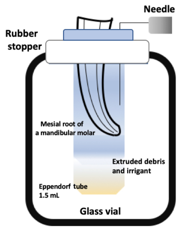
The experimental design defined by Myers and Montgomery (1991) was selected to collect the apically extruded debris. The setup consisted of an Eppendorf tube, a rubber stopper that stabilized the root during preparation and a glass vial (Figure 1). The teeth were placed into the stopper at the CEJ level and fixed with cyanoacrylate Pattex Super Glue (Türk Henkel, Istanbul, Turkey) to prevent solution leakage. The tube into which the debris and irrigants were collected was placed in the glass vial. A 27-gauge needle was placed in the system, within the stopper, to balance internal and external pressure.
Before preparation, the initial weight of the tubes was measured by using an analytical scale (RADWAG, Radom, Poland) with a precision of 10−4 g. Each Eppendorf tube was measured 3 times and the values were averaged. The glass vial was covered with an aluminum leaf to prevent the operator from seeing the apical foramen during preparation.
Root canal preparation
In total, 4 mL of distilled water was used for irrigation in each group during the preparation procedures. All files were used with the same X-Smart Plus endodontic motor (Dentsply Maillefer) according to the manufacturer’s recommendations (Table 1).
Group 1: The OC files were used at 300 rpm speed and 2.5 N·cm torque settings. The path file was used for the initial preparation, and then the OC (25/0.06) files were used for the final preparation.
Group 2: The TRN files were used at 500 rpm speed and 1.5 N·cm torque settings. After the path file, the 20/0.04 (small) and 26/0.04 (prime) files were used for instrumentation.
Group 3: The PTN files were used at 300 rpm speed and 2 N·cm torque settings. The X1 file (17/0.04) was followed by the X2 file (25/0.06) in a brushing outstroke movement.
Evaluation of apical extrusion
The needle, the stopper and the tooth were removed from the tube after the instrumentation was completed. For each specimen, 1 mL of distilled water was used to collect the debris accumulated on the root surface. The distilled water was evaporated in an incubator at 70°C for 5 days to obtain dry debris. The tubes containing the dry debris were re-weighed with the same balance. As in the previous measurement, the samples were weighed 3 times and the values were averaged. The amount of apically extruded debris was calculated by subtracting the weight of the empty tube from the weight of the tube containing the accumulated debris.
Statistical analysis
The statistical analysis employed the IBM SPSS Statistics for Windows software, v. 19.0 (IBM Corp., Armonk, USA). Data was presented as mean and standard deviation (M ±SD). First, the data was analyzed using the Shapiro–Wilk test to verify the assumption of normality. The groups were then compared using the Kruskal–Wallis test for all variables. A p-value of less than 0.05 was considered statistically significant.
Results
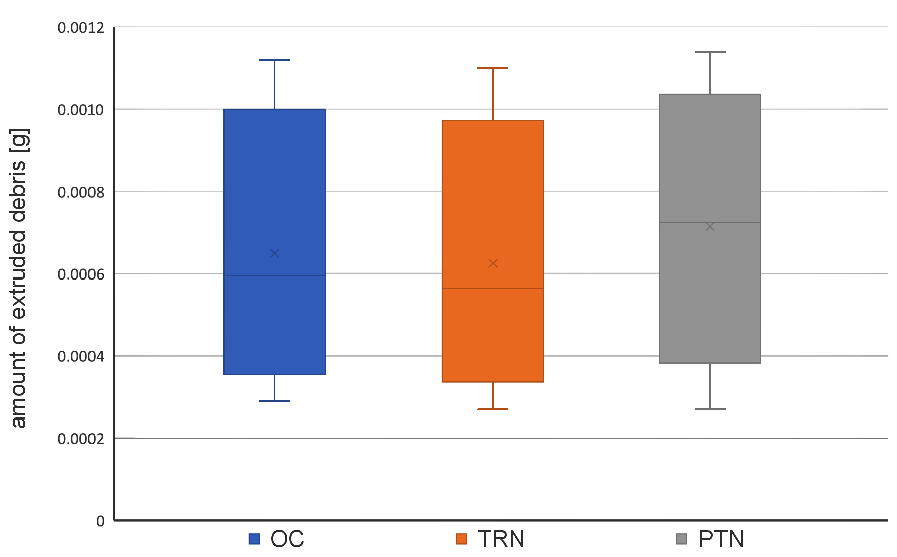
There was no significant difference between the groups (p = 0.270). According to the mean values, the TRN group demonstrated the lowest, while the PTN group demonstrated the highest debris extrusion (p = 0.257). The mean, standard deviation, median, and minimum–maximum values are presented in Table 2 and Figure 2.
Discussion
Apically extruded debris can cause several postoperative complications, such as inflammation and postoperative pain.19 Therefore, a reduction in debris extrusion during root canal treatment may positively affect the postoperative conditions. The preparation system used affects the amount of extruded debris, while the methodology used to collect the debris has a limitation of simulating periapical tissues. According to Versiani et al., a standard comparison can be made by providing the same conditions, which can be achieved in vitro in a laboratory.20 Thus, it may be claimed that the main advantage of the debris collection method used in the present study was standardization.
Distilled water has been proposed as an irrigation solution instead of sodium hypochlorite (NaOCl) so as not to affect the measurements, since the sodium crystals cannot be removed from the debris.21 Tinaz et al. reported that the width of the apical structure could change the results regarding the apically extruded debris.10 To avoid these disadvantages and to standardize the samples in all groups, apical patency was established with a #10 K file. The incidence of postoperative pain due to apical debris extrusion is reportedly higher in curved canals.22, 23 Therefore, in the current study, the mesiobuccal canals of mandibular molar teeth, with moderate to severe curvatures, were selected to compare the 3 different NiTi file systems in terms of apical debris extrusion.
Various NiTi systems are available for the instrumentation of root canals during endodontic treatment, with PTN being a commonly used and accepted system. Various studies have reported the occurrence of debris extrusion after the instrumentation with the use of the PTN files.19, 24 Reddy and Hicks reported that the instrument design was crucial for the amount of apically extruded debris.11 The design of PTN decreases the interaction between the file and dentin, and minimizes the removal of debris from the apex, which is the main advantage of PTN and may cause less apical debris extrusion.21 The PTN files were associated with less extrusion in straight canals as compared to the Controlled Memory NiTi files.24 However, a conflicting result was observed during the instrumentation of curved root canals, with the PTN files associated with significantly more extrusion.25 This discrepancy reveals the relationship between the anatomy of the root and the amount of extruded debris. A micro-computed tomographic evaluation revealed that the PTN system provided suboptimal mechanical preparation for molar teeth and was unable to obtain completely packed debris-free root canal surfaces.26 This conclusion may explain an increase in debris extrusion in posterior teeth, which are relatively difficult to prepare.
The OC system includes a single instrument for preparing the root canal. Reducing the number of files during preparation minimizes the contact area between the file and the dentin wall. Such a situation may be beneficial for reducing the apical extrusion of debris by accommodating more space for the debris around the file.27 However, under the same conditions, no differences were noted between the OC single-file, 2Shape (Micro-Mega) and PTN systems in terms of apical debris extrusion.28 Similarly, Bürklein et al. found no difference between single- and multiple-file systems for the apical extrusion of debris.29 The results of the present study support these findings, as the OC single-file system demonstrated similar results to both the PTN and TRN multiple-rotary file systems. Thus, it can be concluded that the number of files did not significantly affect debris extrusion.
Recently, Tüfenkçi et al. reported that by preparing a contracted endodontic cavity, the OC system caused less apical debris extrusion than the reciprocating single-file system.30 This finding was related to the C-wire heat treatment technology. The C-wire provides the OC files with enhanced flexibility, easy access to canals, and the ability to pre-bend in order to preserve the original form of the root canal during preparation.31, 32 This property may be beneficial for optimal mechanical preparation without unnecessarily removing additional dentin, which might lead to more debris accumulation beyond the apex, especially in curved canals.
The most important difference in the TRN design is the use of 0.8-millimeter NiTi wire instead of the 1.2-millimeter one. The increased flexibility may facilitate the file movement in the root canal during preparation. The special design of the TRN files creates a slim shape that provides more space for debridement,16 while the lower tapered design may help preserve the tooth structure. Limited information is available about the novel TRN system, although cyclic fatigue studies demonstrated promising results for the resistance of the TRN system as compared to various NiTi files.33, 34 The present study demonstrated comparable results for the novel TRN files in relation to the commonly used PTN and OC systems. The lower taper of TRN may prove advantageous, especially in curved canals, by preventing damage to the dentinal structure, and may lead to the reduction of debris extrusion.
Limitations
The main limitation of the present study was that the experimental design could not mimic periapical tissues and their resistance. There are some materials used to imitate their textures. Agar gel and floral foam may be used for the periapical area. However, these materials also have limitations, such as difficulties in setting and achieving definite values for agar gel, and the absorption of the extruded material for the foam.35, 36 Another limitation of this study was the use of extracted teeth for the experiment. The standardization of the extracted root canals was difficult, especially those with curvature. However, using root canals manufactured from acrylic or plastic has several adverse effects. Indeed, the heat generated during preparation might soften the simulated tooth, which could affect the results.37
Within the limitations, our laboratory experiments represent preliminary results for improving the clinical conditions. It is crucial to evaluate the new rotary file systems to minimize apical debris extrusion for the success of endodontic treatment and a comfortable postoperative process.
Conclusions
All instrumentation systems were associated with debris extrusion. The tested file systems presented similar results in terms of apical debris extrusion in curved canals. The novel TRN system demonstrated promising results, comparable OC and PTN.
Ethics approval and consent to participate
The study protocol was accomplished under the Ethics Committee’s standing orders (protocol No. 2020-09-16/18).
Data availability
The datasets generated and/or analyzed during the current study are available from the corresponding author on reasonable request.
Consent for publication
Not applicable.