Dental and Medical Problems

Dent Med Probl
Impact Factor (IF 2024) – 3.9
Journal Citation Indicator (JCI 2024) - 1.36
Scopus CiteScore (2024) – 5.0
Index Copernicus Value (ICV 2023) – 181.00
MNiSW – 70 pts
ISSN 1644-387X (print)
ISSN 2300-9020 (online)
Periodicity – bimonthly


Download original text (EN)

Dental and Medical Problems

2023, vol. 60, nr 1, January-March, p. 153–166

doi: 10.17219/dmp/151482

Publication type: review

Language: English

License: Creative Commons Attribution 3.0 Unported (CC BY 3.0)

Download citation:

  • BIBTEX (JabRef, Mendeley)
  • RIS (Papers, Reference Manager, RefWorks, Zotero)

Cite as:


Pion LA, Matos LLMd, Gimenez T, Palma-Dibb RG, Faraoni JJ. Treatment outcome for dentin hypersensitivity with laser therapy: Systematic review and meta-analysis. Dent Med Probl. 2023;60(1):153–166. doi:10.17219/dmp/151482

Treatment outcome for dentin hypersensitivity with laser therapy: Systematic review and meta-analysis

Luciana Antonio Pion1,A,B,C,D, Laís Lopes Machado De Matos2,A,B,C,D, Thaís Gimenez3,C,E, Regina Guenka Palma-Dibb2,C,D, Juliana Jendiroba Faraoni2,C,E,F

1 Department of Pediatric Dentistry, School of Dentistry of Ribeirão Preto, University of São Paulo, Ribeirão Preto, Brazil

2 Department of Restorative Dentistry, School of Dentistry of Ribeirão Preto, University of São Paulo, Ribeirão Preto, Brazil

3 Department of Pediatric Dentistry, School of Dentistry of São Paulo, University of São Paulo, São Paulo, Brazil

Abstract

Background. Laser protocols for the treatment of dentin hypersensitivity (DH) have not yet been studied systematically.

Objectives. The present study aimed to review clinical trials on the treatment of DH with laser therapy through a systematic review and meta-analysis.

Material and methods. The search of electronic databases resulted in 562 publications up to April 2020. The inclusion criteria were studies carried out on humans and reporting on the treatment of DH with laser therapy. Case reports, literature reviews and systematic reviews were excluded. Selected by abstract, potentially eligible papers were read in full (n = 160). Independent examiners performed data extraction and the assessment of the risk of bias.

Results. A total of 34 studies were included in the analysis, and 11 in the quantitative analysis. It was observed that most studies followed up patients for a maximum of 6 months (55%). Through the meta-analysis, we observed statistically significant differences between the average pain before and after 3 months of treatment with high- and low-power lasers. However, through indirect comparisons, it was observed that the high-power laser showed a greater tendency to reduce the pain levels after 3 months of treatment as compared to the low-power laser, but without a statistically significant difference.

Conclusions. It was possible to conclude that regardless of the type of laser used in the treatment of DH, this treatment is an effective option for the control of pain symptoms. However, it was not possible to establish a defined treatment protocol, since the evaluation methods are very different from each other.

Text for Rewiew and clinical cases.

Keywords: systematic review, laser, hypersensitivity, therapy, dentin

Introduction

Dentin hypersensitivity (DH) is one of the most commonly encountered clinical problems in dental practice. It is defined as pain resulting from exposed dentin in reply to chemical, thermal, tactile, or osmotic stimuli, which cannot be attributed to any other defect or dental pathology.1, 2, 3, 4 Clinically, it is a localized or generalized sharp pain of quick beginning and can affect one or several dental surfaces.5 DH can have an adverse effect on the quality of life, especially concerning limitations of diet, tooth hygiene, and daily activities.6, 7, 8 A significant association was found between DH and the frequency of tooth brushing in a recent study.9 In addition, poor oral hygiene can result in the emergence of other oral diseases and amplification of DH.9 Hypersensitivity in younger patients is often caused by the exposure of dentin due to erosion, while gingival recession often causes hypersensitivity in older patients due to the exposure of the dentinal tubules in the cervical areas caused by periodontal disease and intensified brushing activity.10

Many theories have been proposed to explain the biological mechanism of DH. For example, dentinal receptor mechanisms, odontoblastic transducer mechanisms, pain receptors on nerves in the pulp, nerves stimulated by hydrodynamic mechanisms, and nerve impulses modulated by the release of polypeptides.11 However, the hydrodynamic system theory is the most accepted, and it suggests that the painful sensation is induced by the stimulation of dentin exposed by the movement of fluid within open dentin tubules in reply to mechanical or osmotic alterations/stimuli. The dislocation of fluid in the interior of the tubules causes intrapulpar pressure, which stimulates the nerve fibers located in the dentin-pulp interface and generates tooth pain.12

There is a range of treatment options for DH, such as toothpaste, potassium-based agents, glutaraldehyde-based agents, varnishes, strontium or acetate chlorides, resins, oxalates, calcium phosphates, and laser therapy.13 All procedures work to obliterate the tubules and/or desensitize pulp receptors,14 but none have produced predictable long-term results. This is thought to be because the physical and chemical processes in the oral environment can result in the tubules being exposed again. The ability to resist the recurrence of pain would be essential for laser therapy to maintain its long-term effectiveness.15

However, many studies have shown laser therapy to be an effective treatment for hypersensitivity.16, 17, 18, 19 The treatment can be accomplished with high or low-intensity laser therapies.16, 18, 19, 20, 21 According to the wavelength of the laser, the action on the dentin surface during the treatment of DH is different depending on the irradiation parameters (mainly wavelength and energy density) and also on the time of treatment.22

The first mechanism of action of the high-power laser occurs by the obliteration of tubules through the partial fusion of dentin substrates.6, 23, 24 The low-power laser decreases the chronic inflammatory process present in the cells, by activating the sodium and potassium pumps and obliterating dentin tubules through the formation of tertiary dentin,25 and can promote analgesic, anti-inflammatory, and biomodulatory effects.17, 22 Laser treatment is well tolerated by patients and is considered safe, quick, and painless.26

A systematic review in 2013 by Lin et al. showed that the different desensitizing treatments used in the office had significantly better results than placebo.27 However, these authors did not stratify the results according to the type of laser evaluated. In the same year, another systematic review showed that both lasers – high and low-power – showed effective results in the treatment of DH.16

In contrast, until now, there are no established treatment protocols in the literature for DH with laser therapy and its long-term effectiveness. Considering these facts, this study aimed to review clinical trials and protocols for DH treatment with laser therapy through a systematic review and meta-analysis, in addition to updating the studies available on the proposed subject.

Methods

This article was written using the PRISMA protocol as a guide for the systematic review available at http://www.prisma-statement.org and was registered in the PROSPERO database (CRD42018095822).

Search strategy and selection criteria

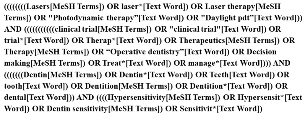
The search was performed and limited to studies in PubMed (MEDLINE) with an English language restriction up to April 2020. The literature search was conducted as displayed in Figure 1.

The selection was conducted by two authors (LAP and LLMM). Each author reviewed the title and abstract of the papers and selected the studies based on inclusion criteria. Disagreements on studies were resolved by a discussion, with the involvement of a third review author when necessary. They were trained to achieve nearly perfect agreement of the Kappa index.

Studies were selected according to the criteria defined below.

The inclusion criteria were: (1) studies carried out on humans; (2) articles that reported on the treatment of DH using laser therapy; and (3) studies that were not case reports, literature reviews, or systematic reviews.

The full text of all eligible articles was subject to the following exclusion criteria: (1) non-randomized clinical trials; (2) not written in the English language; (3) not in permanent teeth; (4) studies without laser therapy treatment; (5) studies that did not have at least 3 months of follow-up after baseline; and (6) studies without sufficient information for data extraction.

Data extraction

The studies that met the inclusion and exclusion criteria were submitted for data extraction. The information was extracted by an author (LAP) and later verified by a second author (LLMM). Both reviewed articles at all stages, and differences were discussed until a consensus was reached.

A standardized data extraction form was used to record the following details:

– study characteristics (author, publication year, title, publication journal, study design, country, setting, funding);

– number of teeth treated;

– participant age;

– study design;

– endpoint assessment of pain;

– follow-up time;

– drop-outs in follow-up;

– mean and standard deviation (M ±SD) values before and after laser therapy treatment – second outcome measures;

– success of intervention judged by pain reduction;

– information about the treatment protocol, such as laser type (high or low-power), application time, number of sessions applied, application range, and laser power and energy – primary outcome measures; and

– adverse events.

If there was not enough information described in the paper, an email was sent to the authors in an attempt to complete the data extraction before it was excluded from the review.

The primary endpoint of interest was to research the protocols existing for DH treatment using laser therapy. The secondary endpoint was to try to verify which DH treatment using laser therapy was more successful through the meta-analysis.

Assessment of the risk of bias

Each selected study was assessed qualitatively using the Cochrane Handbook for the Development of Systematic Intervention Reviews, v. 5.1.0 (Cochrane Handbook). The criteria were divided into main domains associated with randomization, blinding, outcome data, and sample characteristics at baseline. The bias risk assessment was accessed as ‘low risk of bias, ‘high risk of bias, or ‘confuse/unclear’ when it was not possible to identify the information or it was uncertain about the subject or potential bias (classified based on each item of the study criteria). Both reviewers (LAP and JJF) performed independent analyses and disagreement was resolved via a discussion.

Statistical analysis

Direct comparisons were performed between the two treatments using a meta-analysis of random effects. Also, network meta-analyses were conducted to allow indirect comparison between high and low-power lasers. We conducted a Bayesian analysis of mixed treatment comparisons (MTC) to concurrently consider both direct and indirect signs. Studies were performed in similar groups of patients, meeting the assumption of transitivity of this network analysis. First, MTC analyses were computed using both fixed and random models, and the merit of fit for the models was quantified using the residual deviation and the residue information criterion (RIC), with lower scores being better. Because of that, the random effects model with consistent variability among studies was chosen for the final MTC analysis. A node split analysis for unreliability was performed for the pairs with both direct and indirect evidence.

Meta-analyses and network meta-analyses were performed using the R package (GeMTC), v. 3.6.1 (R Foundation for Statistical Computing, Vienna, Austria). For each involved study, mean differences (MDs) and 95% confidence intervals (CIs) were estimated.

Results

Study selection

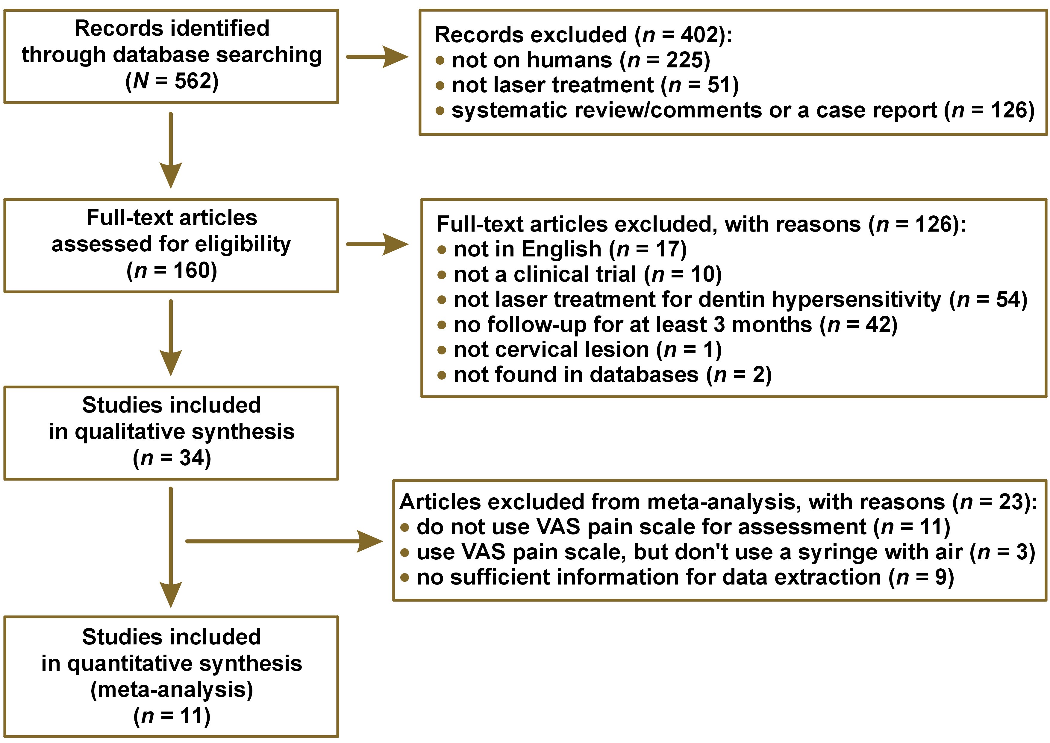
The inter-examiner reproducibility (Kappa index) was 0.85 and considered excellent. The systematic search of the literature identified 562 potentially relevant studies. After removing duplicate studies and screening the titles, abstracts, and/or full texts, 34 studies were included and analyzed qualitatively.

The main reason for not including these studies in the review was that they were not performed on humans (in vivo study, 40%). The remaining 160 studies were analyzed using the full texts for further details and study judgment. Finally, 34 articles matched the eligibility criteria and had adequate data for qualitative analysis, and 11 articles were included in the meta-analysis. The details of the bibliographic research and the selection process of the studies are illustrated in Figure 2.

Study design

The main characteristics of our data sets of longitudinal studies are shown in Table 1.

It was observed that most studies had a split-mouth design (55%), and the age of these subjects ranged from 11 to74 years.

Most of the criteria used for DH assessment and discomfort were reported by the patient. The use of air from a triple syringe followed by the application of a visual probe to scratch the dental surface or perform a thermal test. However, it was also observed that there was no standardization regarding the pain scale used, but most studies use the visual analog scale (VAS) – 70%. The VAS was a 10 cm extension and, with each side, contained an indication of ‘no pain’ and ‘severe pain’.

Follow-up lengths were most frequently 6 months (55%), and only two studies followed the patients for a period of 18 months.

The qualitative analysis included 63 treatment groups, and Table 2 shows the main characteristics of the laser therapy protocols amongst each group, which are described below.

Most of the protocols using lasers with or without another desensitizing agent reduced the pain levels of the patients during the follow-up period. Moreover, the studies that evaluated patients for 18 months had significant results regarding a decrease in pain for all treatments.

The low-power diode laser was the device of choice in 49% of groups that used the low-intensity laser alone or in combination with other therapies. The high-power laser was used alone or combined with other therapies in 55% of the groups that used high-intensity lasers.

It was observed that the average application time for the groups that used a high-power laser was 43 s and that it varied from 1 to 20 applications on the same day. On the other hand, groups using this type of laser did not repeat the treatment protocol on different days, except two studies performed the protocol for three days at intervals of one week per application, and one study performed the protocol on four different days (baseline, 1 week, 1 month, and 6 months). In addition, 31% of the groups evaluated used high-power laser therapy with a desensitizing agent.

In the low-power laser groups, the average application time was 36 s, not exceeding 1 application per day, but being able to repeat applications on different days (maximum 4 applications), except in two groups that performed 4 applications, each lasting 10 s on the same day. Only 9 groups evaluated the use of low-power laser therapy with a desensitizing agent or desensitizing toothpaste.

Risk of bias

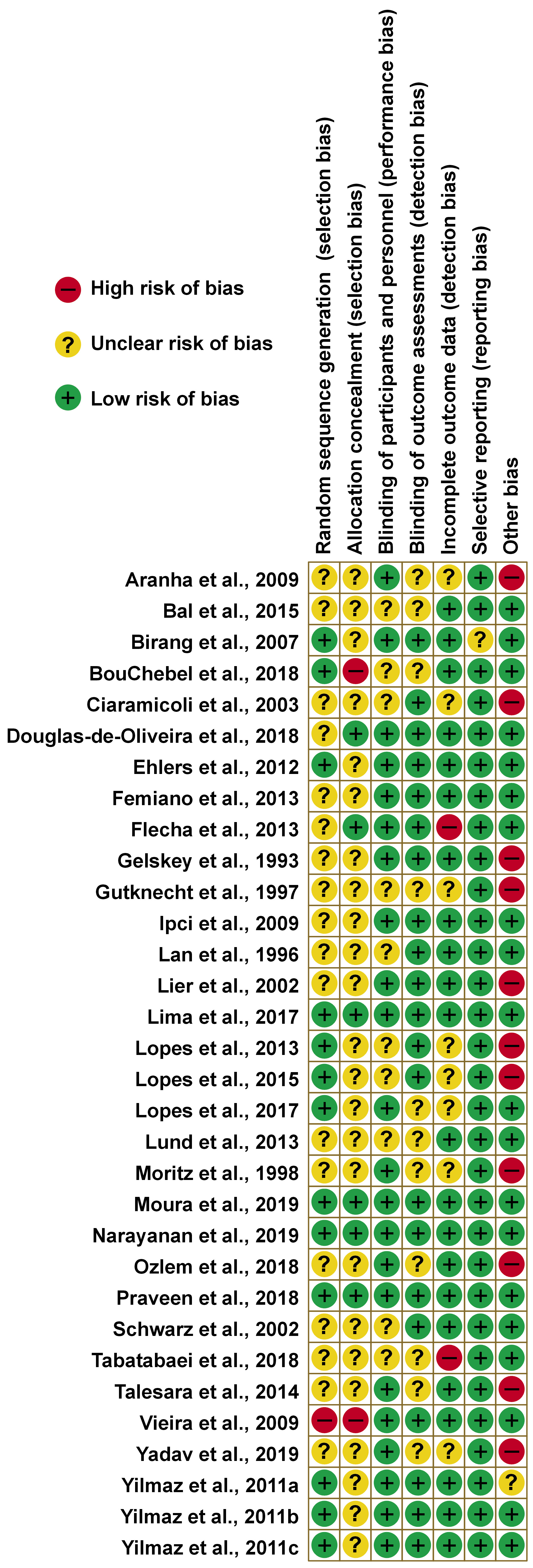
The details of the bias risk assessments for each study are presented in Figure 3. Most studies had one or more “confused/unclear” classifications due to a lack of information. In 40% of the studies, the random sequence generation was described in detail, however, of these, only 4 described the allocation concealment method.

In 68% and 65% of the studies with a low risk of bias had blinding of participants/personnel and outcome assessments, respectively. Regarding incomplete outcome data, the majority had no attrition levels above 20% at the end of the follow-up.

Also, 34% had a high risk of bias concerning the other bias items, as they varied in the number of laser applications administered to the same patient or indicated the use of fluoride-free toothpaste during treatment. In addition, some had a flaw in the methodology that the authors deemed important.

Regarding the articles that were analyzed using the meta-analysis, only 1 did not describe the randomization sequence, 2 did not describe the allocation concealment, and 2 were at high risk due to other biases.

Two studies are not represented in the graph, as they presented only one treatment arm, making it impossible to assess the risk of bias.

Meta-analysis

Only the articles that used the VAS with a syringe of air to assess DH were used for the meta-analysis to standardize the patient’s pain collection. The groups were separated between high and low-intensity lasers using the power described in watts (W) or milliwatts (mW) to perform the meta-analysis separately.

Nine articles were not included in the analysis due to not providing sufficient information about the means and standard deviations before and after treatments. Groups that used desensitizing agents in conjunction with laser therapy were also not included in the meta-analysis.

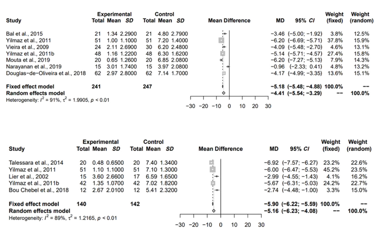
Direct comparisons between the groups

There was a significant difference between average pain levels before and after 3 months of treatment when compared to a treatment control group (before the treatment), regardless of the type of laser used (Figure 4).

Indirect comparisons between the groups

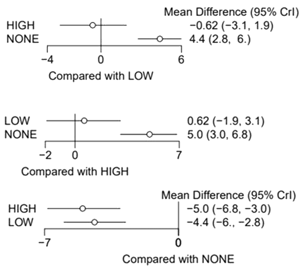
Through indirect comparisons, it was observed that the high-power laser showed a greater tendency to reduce pain levels after 3 months of treatment when compared to the low-power laser, but without a statistically significant difference (Table 3 and Figure 5). Indirect comparisons did not influence the results for low-power versus no treatment and high-power versus no treatment. In that way, there was no inconsistency.

Ranking and network analyses

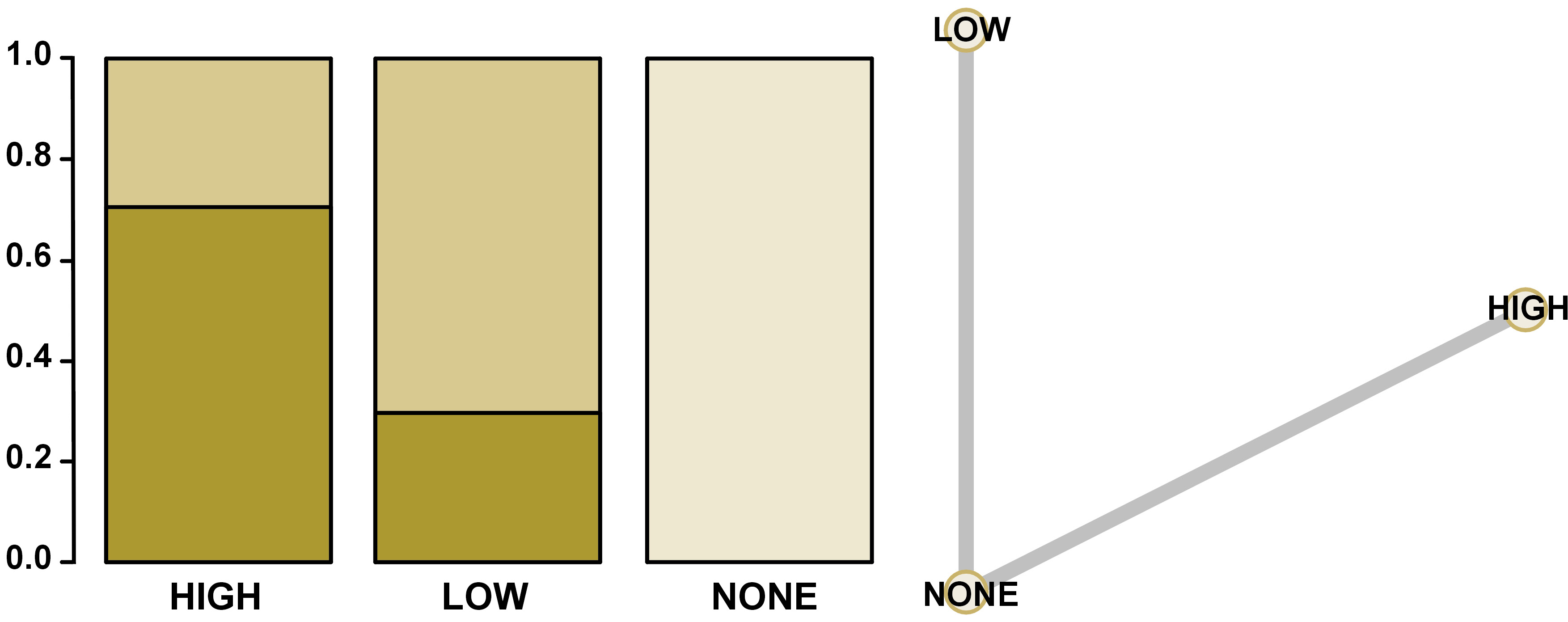
In Figure 6, it was possible to observe through the ranking and network analysis that the high-power lasers showed a greater chance to be the better treatment option, followed by low-power lasers, and performing no treatment the worst concerning self-reported pain for DH. The network diagram is also shown in Figure 6.

Discussion

Various techniques and different protocols for the treatment of DH have been described in the literature, but no systematic reviews and meta-analyses have been performed to date to evaluate DH treatment protocols. A full search strategy was used, though, to decrease the risk of bias, only randomized controlled trials were chosen for this systematic review.

After including the eligible studies, a qualitative analysis of 34 studies was carried out. In contrast, due to the lack of standardization in pain assessments and the lack of information necessary to perform a quantitative analysis, only 11 studies were considered for the meta-analysis.

Through the meta-analysis, it was possible to verify that the average pain reported by patients was lower after 3 months of follow-up when compared to the average pain before treatment with laser therapy.

The management and treatment of DH are complicated as a consequence of their multifactorial nature, and factors such as parafunctional habits, diet, hygiene, and periodontal and pulp health can influence the results, making research about DH complex and challenging.60 Recently, an improvement in laser technology has allowed laser therapy to emerge as a new treatment alternative for DH.61 There are several methods to assess DH intensity, but the lack of standardization in pain measurements is a difficulty faced by researchers.62, 63 This can be explained by the fact that the perception of each individual’s pain is very subjective, which depends directly on the individual’s ability to define what he is feeling through numerical scales.64 The index most used in the studies and evaluated in the qualitative analysis for pain was the VAS (70%).

Various stimuli can be used, such as tactile stimuli using probes and thermal stimuli using a cold water jet and air spray.65 Differences in the mode of DH assessment can also lead to discrepancies in studies, contributing to the heterogeneity of painful responses. Compared to tactile stimulation, air spray is the most reliable method for assessing DH.66 The meta-analysis of this study included only treatment groups that assessed pain by air-jet using a triple syringe, followed by the application of the VAS in an attempt to standardize some parameters to the maximum.

Other variables, such as the wavelength, power, and energy generated by the laser device, are considered of extreme importance since their variability can directly interfere with their applicability. However, it was also observed that these parameters varied among studies, and most high-power laser studies have not described the wavelength and energy used. Which is different from the groups treated with low-power lasers, which described these main parameters. This can be explained since most high-power lasers have pre-established wavelengths depending on the model of the device, and this value can only vary in low-power models.

Split-mouth studies are preferred in dentistry, as it is possible to minimize inter-individual variables, being the participants are their controls, and when these variables are present, they can interfere in the same way with the treatments. However, in cases where it is necessary to assess the patient’s pain on both sides of the mouth, one of the responses may be influenced by the pain the patient is experiencing on the other side of the mouth. It is important to note that most works in this systematic review were carried out using this form.

The low-power lasers used for the treatment of DH can interact with the dental pulp leading to an increase in odontoblastic activity and, consequently, the production of tertiary dentin and a decrease in the lumen size of dentinal tubules. This type of laser can also stimulate cell activity, promote anti-inflammatory effects, and produce analgesic effects.5, 17, 67 High-power lasers, such as carbon dioxide (CO2) and neodymium-doped yttrium aluminum garnet (Nd:YAG), are the typical types of lasers used in dentistry. These high-power lasers can obliterate the dentinal tubules through fusion and re-solidification by melting the hydroxyapatite and, after cooling, solidifying and forming hydroxyapatite crystals larger than in the initial structure, thus reducing DH.44, 68

Most of the studies evaluated, regardless of the type of laser used, showed satisfactory results regarding DH pain after months of therapy. The low-power laser most used by the authors was the diode laser. It is known that, in addition to having an immediate analgesic effect, it stimulates odontoblasts in the production of irregular dentin that can lead to the obliteration of dentinal tubules,69, 70 and provide analgesia after months of treatment.

Some authors have reported the disadvantages of lasers, including that they are very expensive, intricate to use, and use over time decreases, among others.5 However, new research shows that laser therapy is well tolerated by patients and is safe and effective.26, 38 After performing the meta-analysis of treatments with high and low-power lasers, it was observed that both lasers showed a reduction in DH after 3 months of treatment by alleviating symptoms through the formation of reactionary dentin after irradiation.52, 71

In this work, through indirect comparisons, it was found that the high-power laser had lower average pain scores after 3 months of treatment when compared to the low-power laser or with the average pain before treatments, however, there was no difference between them. But, management for DH with laser therapy in the office constitutes a more expensive and elaborate treatment modality than traditional treatment planning,5 and high-power equipment has a higher cost when compared to low-power equipment. Therefore, as the results of high and low-power lasers are similar, the low lasers seem to be a viable option concerning the cost for the clinician, such as the diode laser. Besides that, no study has evaluated their cost-effectiveness and more long-term studies are needed.

As a limitation of this study, it can be observed that the DH assessment methods are very different among studies, generating heterogeneous responses concerning pain, not only because the instrument of each study is different, but also because pain is a subjective and personal experience. In addition, some studies did not report the complete protocol used for each type of laser, often not being possible to include them in the analyses. Although most studies followed patients for 6 months after treatment (55%), it was decided to perform the meta-analysis with a 3-month follow-up to include more treatment groups in the quantitative analysis.

Regardless of the type of laser used in the treatment of DH, laser therapy is a proven choice for the control of pain symptoms, but it is not possible to establish a defined treatment protocol since the evaluations of each method are very different among the studies in this review. Also, the available signs suggest that high and low-power lasers are effective in decreasing DH after 3 months of treatment.

Conclusions

Laser therapy can be an effective choice for the control of pain symptoms, despite the different lasers and protocols studied for HD. According to the studies, it is suggested that lasers are effective in decreasing DH pain after 3 months of treatment.

Ethics approval and consent to participate

Not applicable.

Data availability

The datasets and all supplementary materials generated and/or analyzed during the current study are available from the corresponding author on reasonable request.

Consent for publication

Not applicable.

Tables


Table 1. Characteristics of the clinical trials on the laser therapy for dental hypersensivity (DH) selected for qualitative analysis (­ = 34)

Author
(year)

Number
of teeth

Participant
age
[years]

Study design

Treatment protocol

End point
Assessment of pain

Follow-up
time
[months]

Drop-outs
(%)

Moura et al.
(2019)28

60

18

Parallel groups

-GaAIAS laser

Air stimulus + VAS scale

6

0

El Mobadder et al.
(2019)29

Not reported

Not reported

Single arm

- Diode laser

Air stimulus +water + VAS scale

6

3.2

Yadav et al.
(2019)30

Not reported

Not reported

Parallel groups

- Nanocrystalline hydroxypatite + Nd:YAG + scaling and root planning (SRP)
- SRP + Nd:YAG + herbal dentifrice
- SRP + Nd:YAG

Air stimulus +water + VRS scale

6

Not reported

Narayanan et al.
(2019)31

Not reported

18-60

Parallel groups

- Diode laser
- Diode laser + potassium nitrate desensitising paste

Air + thermal + tactile stimulus + VAS scale

3

0

Tabatabaei et al.
(2018)32

135

25-58

Parallel groups

- Diode laser
- Nd:YAG

Thermal and not reported scale

6

0

BouChebel et al.
(2018)33

54

20-60

Split-mouth

- Nd:YAG

Air, tactile, thermal stimulus + VAS scale

6

0

Praveen et al.
(2018)34

50

20-50

Parallel groups

- Diode laser

Air + water stimulus + VAS scale

3

0

Douglas-de-Oliveira et al.
(2018)35

434

12-60

Split-mouth

- GaAIAS laser

Air stimulus + VAS scale

6

0

Ozlem et al.
(2018)36

100

18-56

Parallel groups

- Nd:YAG
- Gluma desensitizer (DG) + Nd:YAG
- Er,Cr:YSGG
- DG + Er,Cr:YSGG

Probe (Tactilstimulus) + Pain yes or no

6

0%

Lopes et al.
(2017)17

117

22-53

Parallel groups

- Low-power laser, low parameters (LPLD)
- Low-power laser, high parameters (LPHD)
- LPLD + DG
- LPHD + DG
- Nd:YAG laser
- Nd:YAG laser + DG
- LPLD + Nd:YAG
- LPHD + Nd:YAG

Air stimulus + probe + VAS scale

18

Not reported

Lima et al.
(2017)37

434

12-60

Slipt-mouth

- Diode laser

Air stimulus + VAS scale

3

0%

Lopes et al.
(2015)38

55

22-53

Parallel groups

- LPLD
- LPHD
- LPLD + DG
- LPHD + DG

Air stimulus + VAS scale

6

0%

Bal et al.
(2015)39

156

19-60

Slipt-mouth

- Diode laser
- Diode laser + Sensitivity paste
- Sensitivity paste + Diode laser

Air stimulus + VAS scale

6

0%

Talesara et al.
(2014)40

80

25-55

Slipt-mouth

- Nd:YAG laser

Air stimulus + thermal test + VAS scale

9

0%

Femiano et al.
(2013)41

262

21-64

Slipt-mouth

- Diode laser
- NaF + Diode laser

Air stimulus + VAS scale

6

0%

Lund et al.
(2013)42

117

19-58

Slipt-mouth

- Diode laser

Air stimulus + VAS scale

3

0%

Flecha et al.
(2013)43

432

Not reported

Slipt-mouth

-GaAlAs laser

Air stimulus + number scale

6

0%

Lopes et al.
(2013)44

33

Not reported

Split-mouth

-Nd:YAG laser
-Nd:YAG laser + DG

Air stimulus + probe + VAS scale

6

0%

Ehlers et al.
(2012)45

181

Not reported

Split-mouth

- Er,Cr:YSGG laser

Air stimulus + VAS scale

6

21%

Yilmaz et al.
(2011)25

174

18-60

Split-mouth

-Diode laser
- Er,Cr:YSGG laser

Air stimulus + VAS scale

3

0%

Yilmaz et al.
(2011)46

48

11-58

Split-mouth

-GaAlAs laser

Air stimulus + VAS scale

6

0%

Yilmaz et al.
(2011)47

146

Not reported

Split-mouth

- Er, Cr: YSGG

Air stimulus + VAS scale

3

0%

Vieira et al.
(2009)48

164

24-68

Parallel groups

- Diode laser

Air stimulus + probe + VAS scale

3

20%

Ipci et al.
(2009)49

420

23-62

Parallel groups

- CO2 laser
- ER: YAG laser
- CO2 laser + NaF
- ER:YAG laser+ NaF

Air stimulus + Ushida method

6

0%

Aranha et al.
(2009)50

101

Not reported

Parallel groups

- Diode laser

Air stimulus + VAS scale

6

Not reported

Birang et al.
(2007)51

63

Not reported

Split-mouth

- Nd:YAG laser
- Er:YAG laser

Probe + VAS scale

6

0%

Ciaramicoli et al.
(2003)52

145

23-63

Parallel groups

-Nd:YAG laser

Air stimulus + probe + pain scale

6

Not reported

Lier et al.
(2002)53

34

26-66

Split-mouth

-Nd:YAG laser

Air stimulus + VAS scale

4

0%

Schwarz et al.
(2002)54

208

23-56

Split-mouth

- Er:YAG laser

Air stimulus + Pain scale (4 degrees)

6

0%

Zhang et al.
(1998)55

91

25-74

Single arm

-CO2 laser

Air stimulus + pain scale (3 degrees)

3

0%

Moritz et al.
(1998)56

144

Mean 37-45

Parallel groups

-CO2 laser + strontium chloride

Air stimulus + probe + Pain scale (4 degrees)

18

Not reported

Gutknecht et al.
(1997)57

120

26-62

Split-mouth

-Nd:YAG laser

Air stimulus + probe + Pain scale (3 degrees)

3

Not reported

Lan et al.
(1996)58

34

26-66

Split-mouth

-Nd:YAG laser

Air stimulus + VAS scale

4

11%

Gelskey et al.
(1993)59

19

25-69

Split-mouth

-Helium neon laser
-Helium neon laser + Nd:YAG laser

Mechanical and thermal stimulus + VAS scale

3

5%

GaAlAs – gallium-aluminum-arsenide; Nd:YAG – neodymium-doped yttrium aluminum garnet; SRP – scaling and root planing; DG – Gluma desensitizer; Er,Cr:YSGG – erbium, chromium-doped yttrium, scandium, gallium garnet; LPLD – low-power laser, low parameters; LPHD – low-power laser, high parameters; NaF – fluoride sodium; CO2 – carbon dioxide; VAS – visual analog scale.
Table 2. Main features of the laser therapy protocols for each group (n = 51)

Author (year)

Laser
type

Potency

Potency [W/mW]

Energy density
[J/cm2]

Wavelength [nm]

Application time [s]

Same-day sessions

Application interval time on different days

Number of repetitions on different days

Comments

Yadav
et al. (2019)30

Nanocrystalline hydroxyapatite + Nd:YAG + SRP

High

1W

Not
reported

Not
reported

60

5to 20

1 week, 1 month, 6 months

4

Nano crystalline hydroxyapatite 2x/day

Yadav
et al. (2019)30

SRP + Nd:YAG + herbal dentifrice

High

1W

Not
reported

Not
reported

60

5to 20

1 week, 1 month, 6 months

4

Herbal dentifrice 2x/day

Yadav
et al. (2019)30

SRP + Nd:YAG

High

1W

Not
reported

Not
reported

60

5to 20

1 week, 1 month, 6 months

4

-

Tabatabaei
et al. (2018)32

Nd:YAG

High

1W

Not
reported

1064

30

1

1 week

3

-

BouChebel
et al. (2018)33

Nd:YAG

High

0.64W

100

Not
reported

20

4

-

1

-

Ozlem
et al. (2018)36

Nd:YAG

High

1W

35.8

Not
reported

20

3

-

1

-

Ozlem
et al. (2018)36

DG + Nd:YAG

High

1W

35.8

Not
reported

20

3

-

1

30-60sec DG application after laser therapy

Ozlem
et al. (2018)36

Er,Cr:YSGG

High

0.25W

44.3

Not
reported

30

1

-

1

Ozlem
et al. (2018)36

DG + Er,Cr:YSGG

High

0.25W

44,3

Not
reported

30

1

-

1

30-60sec DG application after laser therapy

Lopes
et al. (2017)17

Nd:YAG

High

1W

85

Not
reported

15

4

-

1

-

Lopes
et al. (2017)17

Nd:YAG + DG

High

1W

85

Not
reported

15

4

-

1

30-60sec DG application after 3rd laser therapy

Talesara
et al. (2014)40

Nd:YAG

High

1W

Not
reported

Not
reported

60

2

-

1

-

Lopes
et al. (2013)44

Nd:YAG

High

1.5W

85

Not
reported

60

4

-

1

-

Lopes
et al. (2013)44

Nd:YAG + DG

High

1.5W

85

Not
reported

60

4

-

1

30-60sec DG application after 3rd laser therapy

Ehlers
et al. (2012)45

Er,Cr:YSGG

High

0.001W

0.06

665

Not
reported

1

-

1

-

Yilmaz
et al. (2011)25

Er,Cr:YSGG

High

0.25W

Not
reported

2780

30

1

-

1

-

Yilmaz
et al. (2011)47

Er,Cr:YSGG

High

0.25W

Not
reported

Not
reported

30

1

-

1

-

Ipci
et al. (2009)49

CO2 laser

High

1W

Not
reported

Not
reported

10

1

-

1

-

Ipci
et al. (2009)49

Er:YAG laser

High

Not
reported

60

Not
reported

10

1

-

1

-

Ipci
et al. (2009)49

CO2 laser + NaF

High

1W

Not
reported

Not
reported

10

1

-

1

NaF applied for 4 min

Ipciet al. (2009)49

Er:YAG laser + NaF

High

Not
reported

60

Not
reported

10

1

-

1

NaF applied for 4 min

Birang
et al. (2007)51

Er:YAG laser

High

Not
reported

100

Not
reported

60

2

-

1

-

Birang
et al. (2007)51

Nd:YAG laser

High

1W

Not
reported

Not
reported

60

2

-

1

-

Ciaramicoli
et al. (2003)52

Nd:YAG

High

1W

0.04

1064

30

1

1 week

3

-

Lier
et al. (2002)53

Nd:YAG laser

High

4W

Not
reported

Not
reported

120

1

-

1

-

Schwarz
et al. (2002)54

Er:YAG laser

High

Not
reported

80

Not
reported

120

1

-

1

-

Zhag
et al. (1998)55

CO2 laser

High

1W

Not
reported

Not
reported

0,5

3

-

1

-

Moritz
et al. (1998)56

CO2 laser + strontium chloride

High

0.5W

Not
reported

Not
reported

5

6

-

1

The number of laser applications varies according to the pain reported by the patient

Gutknecht
et al. (1997)57

Nd:YAG laser

High

0.3W

Not
reported

1064

30-90

1

-

1

-

Gutknecht
et al. (1997)57

Nd:YAG laser

High

0.6W

Not
reported

1064

30-90

1

-

1

-

Gutknecht
et al. (1997)57

Nd:YAG laser

High

1W

Not
reported

1064

30-90

1

-

1

-

Lan
et al. (1996)58

Nd:YAG laser

High

Not
reported

30

Not
reported

120

1

-

1

-

Moura
et al. (2019)28

GAIAs laser

Low

100mW

4

808

40

4

48h

4

-

El Mobadder
et al. (2019)29

Diode laser

Low

1000mW

Not
reported

980

Variable

Not
reported

-

1

-

Narayanan
et al. (2019)31

Diode laser

Low

1000mW

Not
reported

810

10

Not
reported

Not
reported

Not
reported

-

Narayanan
et al. (2019)31

Diode laser + potassium nitrate

Low

1000mW

Not
reported

810

10

Not
reported

Not
reported

Not
reported

-

Tabatabaei
et al. (2018)32

Diode laser

Low

200mW

84.4

810

30

1

1 week

3

-

Praveen
et al. (2018)34

Diode laser

Low

60mW

9

905

60

1

-

1

-

Douglas-de-Oliveira
et al. (2018)35

GaAIAS laser

Low

120mW

30.9

795

8

1

48h

3

-

Lopes
et al. (2017)17

LPLD

Low

30mW

10

Not
reported

9

1

72h

3

-

Lopes
et al. (2017)17

LPHD

Low

100mW

40

Not
reported

11

1

72h

3

-

Lopes
et al. (2017)17

LPLD + DG

Low

30mW

10

Not
reported

9

1

72h

3

30-60sec DG application after 3rd laser therapy

Lopes
et al. (2017)17

LPHD + DG

Low

100mW

40

Not
reported

11

1

72h

3

30-60sec DG application after 3rd laser therapy

Lima
et al. (2017)37

Diodo laser

Low

120mW

30.96

795

8

1

48h

3

-

Lopes
et al. (2015)38

LPLD

Low

30mW

10

635-685

9

1

72h

3

-

Lopes
et al. (2015)38

LPHD

Low

100mW

90

808-830

11

1

72h

3

-

Lopes
et al. (2015)38

LPLD + DG

Low

30mW

10

635-685

9

1

72h

3

30-60sec DG application after 3rd laser therapy

Lopes
et al. (2015)38

LPHD + DG

Low

100mW

90

808-830

11

1

72h

3

30-60sec DG application after 3rd laser therapy

Bal
et al. (2015)39

Diodo laser

Low

25mW

2

685

100

1

-

1

-

Bal
et al. (2015)39

Diodo laser+ toothpaste for sensitivity

Low

25mW

2

685

100

1

-

1

Applying the toothpaste after laser therapy 2 times one after the other

Balet al. (2015)39

Toothpaste for sensitivity+ Diodo laser

Low

25mW

2

685

100

1

-

1

Applying the toothpaste after laser therapy 2 times one after the other

Femiano
et al. (2013)41

Diode laser

Low

200mW

-

808

60

1

168h

3

-

Femiano
et al. (2013)41

NaF + Diode laser

Low

200mW

-

808

60

1

168h

3

NaF applied for 60 sec

Lund
et al. (2013)42

Diode laser

Low

20mW

5

780

40

4

72h

3

-

Flecha
et al. (2013)43

GAIAs laser

Low

120mW

2.88

785

8

1

48h

3

-

Yilmaz
et al. (2011)46

GAIAs laser

Low

500mW

5

810

60

1

-

1

-

Yilmaz
et al. (2011)25

Diodo laser

Low

Not
reported

8.5

810

60

1

-

1

-

Vieira
et al. (2009)48

Diodo laser

Low

30mW

4

660

120

1

168h

4

-

Aranha
et al. (2009)50

Diodo laser

Low

15mW

3.8

660

10

1

72h

3

-

Gelskey
et al. (1993)59

Helium neon laser

Low

Not
reported

30

632

10

Varies

-

1

The number of laser applications varies according to the pain reported by the patient

Lopes
et al. (2017)17

LPLD + Nd:YAG

Low/high

30mW/1W

10/85

635-685/400000

9/15

¼

72h/0

3/1

Nd:YAG laser application on 3rd session

Lopes
et al. (2017)17

LPHD + Nd:YAG

Low/high

100mW/1W

40/85

808-830/400000

11/15

¼

72h/0

3/1

Nd:YAG laser application on 3rd session

Gelskey
et al. (1993)59

Helium neon laser + Nd:YAG laser

Low/high

Not
reported

30

632/1064

10

Varies

-

1

The number of laser applications varies according to the pain reported by the patient

Table 3. Indirect comparisons between the groups

Direct evidence
MD (95% CI)

MTC (direct + indirect evidence)
MD (95% CI)

High vs. none

5.0 (3.0–6.8)

5.0 (3.0–6.8)

Low vs. none

4.4 (2.8–6.0)

4.4 (2.8–6.0)

High vs. low

0.62 (−1.9–3.1)

MD – mean difference; CI – confidence interval; MTC – mixed treatment comparison: high laser, none – before the treatment, low laser.

Figures


Fig. 1. Search strategy
Fig. 2. PRISMA flowchart of the literature search, determination of eligibility, and inclusion in the systematic review and meta-analysis
Fig. 3. Methodological quality and the risk of bias of longitudinal clinical trials selected for the study
Red, green and yellow colors refer to a high risk of bias, a low risk of bias and an unclear risk of bias, respectively.
Fig. 4. Forest plot of comparison: Pain reported by the patient before and after 3 months of treatment with a low-power laser and a high-power laser respectively
Fig. 5. Forest plot of indirect comparisons between the groups
Fig. 6. Ranking and network analyses

References (71)

  1. Addy M. Etiology and clinical implications of dentine hypersensitivity. Dent Clin North Am. 1990;34(3):503–514. PMID:2197124.
  2. Holland GR, Narhi MN, Addy M, Gangarosa L, Orchardson R. Guidelines for the design and conduct of clinical trials on dentine hypersensitivity. Journal of clinical periodontology. Nov 1997;24(11):808-813. doi:10.1111/j.1600-051x.1997.tb01194.x
  3. West NX. Dentine hypersensitivity. Monographs in oral science. 2006;20:173-189. doi:10.1159/000093362
  4. Felix J, Ouanounou A. Dentin Hypersensitivity: Etiology, Diagnosis, and Management. Compendium of continuing education in dentistry (Jamesburg, NJ : 1995). Nov/Dec 2019;40(10):653-657; quiz 658. PMID:31730363.
  5. Orchardson R, Gillam DG. Managing dentin hypersensitivity. Journal of the American Dental Association. Jul 2006;137(7):990-8; quiz 1028-9. doi:10.14219/jada.archive.2006.0321
  6. Davari A, Ataei E, Assarzadeh H. Dentin hypersensitivity: etiology, diagnosis and treatment; a literature review. Journal of dentistry (Shiraz, Iran). Sep 2013;14(3):136-45. PMID:24724135. PMCID:PMC3927677.
  7. Idon PI, Sotunde OA, Ogundare TO. Beyond the Relief of Pain: Dentin Hypersensitivity and Oral Health-Related Quality of Life. Frontiers in dentistry. Sep-Oct 2019;16(5):325-334. doi:10.18502/fid.v16i5.2272
  8. West NX. Dentine hypersensitivity: preventive and therapeutic approaches to treatment. Periodontology 2000. 2008;48:31-41. doi:10.1111/j.1600-0757.2008.00262.x
  9. Blaizot A, Offner D, Trohel G, et al. Prevalence of sensitive teeth and associated factors: a multicentre, cross-sectional questionnaire survey in France. BMC oral health. Aug 26 2020;20(1):234. doi:10.1186/s12903-020-01216-1
  10. Splieth CH, Tachou A. Epidemiology of dentin hypersensitivity. Clin Oral Investig. Mar 2013;17 Suppl 1(Suppl 1):S3-8. doi:10.1007/s00784-012-0889-8
  11. West N, Seong J, Davies M. Dentine hypersensitivity. Erosive Tooth Wear. 2014;25:108-122. doi:10.1159/000360749
  12. Brannstrom M. The hydrodynamic theory of dentinal pain: Sensation in preparations, caries, and the dentinal crack syndrome. J Endod. 1986;12(10):453–457. doi:10.1016/S0099-2399(86)80198-4
  13. Zeola LF, Teixeira DNR. Brazilian dentists’ perception of dentin hypersensitivity management. 2020;33:e115. doi:10.1590/1807-3107bor-2019.vol33.0115
  14. Figueiredo Macedo de Lima J, Aguiar Jordão Mainardi MDC, Puppin-Rontani RM, et al. Bioinspired catechol chemistry for dentin remineralization: A new approach for the treatment of dentin hypersensitivity. Dental materials : official publication of the Academy of Dental Materials. Apr 2020;36(4):501-511. doi:10.1016/j.dental.2020.01.012
  15. Machado AC, Viana Í EL, Farias-Neto AM, et al. Is photobiomodulation (PBM) effective for the treatment of dentin hypersensitivity? A systematic review. Lasers in medical science. May 2018;33(4):745-753. doi:10.1007/s10103-017-2403-7
  16. Sgolastra F, Petrucci A, Severino M, Gatto R, Monaco A. Lasers for the treatment of dentin hypersensitivity: a meta-analysis. Journal of dental research. Jun 2013;92(6):492-9. doi:10.1177/0022034513487212
  17. Lopes AO, de Paula Eduardo C, Aranha ACC. Evaluation of different treatment protocols for dentin hypersensitivity: an 18-month randomized clinical trial. Lasers in medical science. Jul 2017;32(5):1023-1030. doi:10.1007/s10103-017-2203-0
  18. Lee SY, Jung HI, Jung BY, Cho YS, Kwon HK, Kim BI. Desensitizing efficacy of nano-carbonate apatite dentifrice and Er,Cr:YSGG laser: a randomized clinical trial. Photomedicine and laser surgery. Jan 2015;33(1):9-14. doi:10.1089/pho.2014.3787
  19. Femiano F, Femiano R, Femiano L, et al. A new combined protocol to treat the dentin hypersensitivity associated with non-carious cervical lesions: A randomized controlled trial. Appl Sci. 2020;11(1):187. doi:10.3390/app11010187
  20. Shiau HJ. Dentin hypersensitivity. The journal of evidence-based dental practice. Sep 2012;12(3 Suppl):220-8. doi:10.1016/s1532-3382(12)70043-x
  21. Palazon MT, Scaramucci T, Aranha AC, et al. Immediate and short-term effects of in-office desensitizing treatments for dentinal tubule occlusion. Photomedicine and laser surgery. Jun 2013;31(6):274-82. doi:10.1089/pho.2012.3405
  22. Kimura Y, Wilder-Smith P, Yonaga K, Matsumoto K. Treatment of dentine hypersensitivity by lasers: a review. Journal of clinical periodontology. Oct 2000;27(10):715-21. doi:10.1034/j.1600-051x.2000.027010715.x
  23. Umana M, Heysselaer D, Tielemans M, Compere P, Zeinoun T, Nammour S. Dentinal tubules sealing by means of diode lasers (810 and 980 nm): a preliminary in vitro study. Photomedicine and laser surgery. Jul 2013;31(7):307-14. doi:10.1089/pho.2012.3443
  24. Rosa RR, Calazans FK, Nogueira RD, Lancellotti AC, Gonçalves LS, Geraldo-Martins VR. Effects of different desensitizing treatments on root dentin permeability. Brazilian oral research. Oct 10 2016;30(1):e111. doi:10.1590/1807-3107BOR-2016.vol30.0111
  25. Yilmaz HG, Kurtulmus-Yilmaz S, Cengiz E, Bayindir H, Aykac Y. Clinical evaluation of Er,Cr:YSGG and GaAlAs laser therapy for treating dentine hypersensitivity: A randomized controlled clinical trial. Journal of dentistry. Mar 2011;39(3):249-54. doi:10.1016/j.jdent.2011.01.003
  26. Hashim NT, Gasmalla BG, Sabahelkheir AH, Awooda AM. Effect of the clinical application of the diode laser (810 nm) in the treatment of dentine hypersensitivity. BMC research notes. Jan 13 2014;7:31. doi:10.1186/1756-0500-7-31
  27. Lin PY, Cheng YW, Chu CY, Chien KL, Lin CP, Tu YK. In-office treatment for dentin hypersensitivity: a systematic review and network meta-analysis. Journal of clinical periodontology. Jan 2013;40(1):53-64. doi:10.1111/jcpe.12011
  28. Moura GF, Zeola LF, Silva MB, Sousa SC, Guedes FR, Soares PV. Four-Session Protocol Effectiveness in Reducing Cervical Dentin Hypersensitivity: A 24-Week Randomized Clinical Trial. Photobiomodulation, photomedicine, and laser surgery. Feb 2019;37(2):117-123. doi:10.1089/photob.2018.4477
  29. El Mobadder M, Namour A, Namour M, et al. Dentinal hypersensitivity treatment using diode laser 980 nm: In vivo study. Dent J (Basel). 2019;7(1):5. doi:10.3390/dj7010005
  30. Yadav RK, Verma UP, Tiwari R. Comparative evaluation of neodymium-doped yttrium aluminum garnet laser with nanocrystalline hydroxyapatite dentifrices and herbal dentifrices in the treatment of dentinal hypersensitivity. National journal of maxillofacial surgery. Jan-Jun 2019;10(1):78-86. doi:10.4103/njms.NJMS_47_18
  31. Narayanan R, Prabhuji MLV, Paramashivaiah R, Bhavikatti SK. Low-level Laser Therapy in Combination with Desensitising Agent Reduces Dentin Hypersensitivity in Fluorotic and Non-fluorotic Teeth - A Randomised, Controlled, Double-blind Clinical Trial. Oral Health Prev Dent. 2019;17(6):547-556. doi:10.3290/j.ohpd.a43567
  32. Tabatabaei MH, Chiniforush N, Hashemi G, Valizadeh S. Efficacy Comparison of Nd:YAG laser, diode laser and dentine bonding agent in dentine hypersensitivity reduction: a clinical trial. Laser therapy. Dec 31 2018;27(4):265-270. doi:10.5978/islsm.27_18-OR-24
  33. Bou Chebel F, Zogheib CM, Baba NZ, Corbani KA. Clinical Comparative Evaluation of Nd:YAG Laser and a New Varnish Containing Casein Phosphopeptides-Amorphous Calcium Phosphate for the Treatment of Dentin Hypersensitivity: A Prospective Study. Journal of prosthodontics: official journal of the American College of Prosthodontists. Dec 2018;27(9):860-867. doi:10.1111/jopr.12984
  34. Praveen R, Thakur S, Kirthiga M, Narmatha M. Comparative evaluation of a low-level laser and topical desensitizing agent for treating dentinal hypersensitivity: A randomized controlled trial. Journal of conservative dentistry: JCD. Sep-Oct 2018;21(5):495-499. doi:10.4103/jcd.jcd_197_18
  35. Douglas-de-Oliveira DW, Lages FS, Ramos-Jorge ML, Ramos Glória JC, Gonçalves PF, Flecha OD. Laser and cyanoacrylate for the treatment of dentine hypersensitivity: Survival analysis and predictive factors. J Int Acad Periodontol. 2018;20(3):102–109. PMID:31522166.
  36. Ozlem K, Esad GM, Ayse A, Aslihan U. Efficiency of Lasers and a Desensitizer Agent on Dentin Hypersensitivity Treatment: A Clinical Study. Nigerian journal of clinical practice. Feb 2018;21(2):225-230. doi:10.4103/njcp.njcp_411_16
  37. Lima TC, Vieira-Barbosa NM, Grasielle de Sá Azevedo C, et al. Oral Health-Related Quality of Life Before and After Treatment of Dentin Hypersensitivity With Cyanoacrylate and Laser. Journal of periodontology. Feb 2017;88(2):166-172. doi:10.1902/jop.2016.160216
  38. Lopes AO, Eduardo Cde P, Aranha AC. Clinical evaluation of low-power laser and a desensitizing agent on dentin hypersensitivity. Lasers in medical science. Feb 2015;30(2):823-9. doi:10.1007/s10103-013-1441-z
  39. Bal MV, Keskiner İ, Sezer U, Açıkel C, Saygun I. Comparison of low level laser and arginine-calcium carbonate alone or combination in the treatment of dentin hypersensitivity: a randomized split-mouth clinical study. Photomedicine and laser surgery. Apr 2015;33(4):200-5. doi:10.1089/pho.2014.3873
  40. Talesara K, Kulloli A, Shetty S, Kathariya R. Evaluation of potassium binoxalate gel and Nd:YAG laser in the management of dentinal hypersensitivity: a split-mouth clinical and ESEM study. Lasers in medical science. Jan 2014;29(1):61-8. doi:10.1007/s10103-012-1239-4
  41. Femiano F, Femiano R, Lanza A, Festa MV, Rullo R, Perillo L. Efficacy of diode laser in association to sodium fluoride vs Gluma desensitizer on treatment of cervical dentin hypersensitivity. A double blind controlled trial. Am J Dent. 2013;26(4):214–218. PMID:24693632.
  42. Lund RG, Silva AF, Piva E, Da Rosa WL, Heckmann SS, Demarco FF. Clinical evaluation of two desensitizing treatments in southern Brazil: A 3-month follow-up. Acta odontologica Scandinavica. Nov 2013;71(6):1469-74. doi:10.3109/00016357.2013.770919
  43. Flecha OD, Azevedo CG, Matos FR, et al. Cyanoacrylate versus laser in the treatment of dentin hypersensitivity: a controlled, randomized, double-masked and non-inferiority clinical trial. Journal of periodontology. Mar 2013;84(3):287-94. doi:10.1902/jop.2012.120165
  44. Lopes AO, Aranha AC. Comparative evaluation of the effects of Nd:YAG laser and a desensitizer agent on the treatment of dentin hypersensitivity: a clinical study. Photomedicine and laser surgery. Mar 2013;31(3):132-8. doi:10.1089/pho.2012.3386
  45. Ehlers V, Ernst CP, Reich M, Kämmerer P, Willershausen B. Clinical comparison of gluma and Er:YAG laser treatment of cervically exposed hypersensitive dentin. Am J Dentistry. 2012;25(3):131–135. PMID:22988681.
  46. Yilmaz HG, Kurtulmus-Yilmaz S, Cengiz E. Long-term effect of diode laser irradiation compared to sodium fluoride varnish in the treatment of dentine hypersensitivity in periodontal maintenance patients: a randomized controlled clinical study. Photomedicine and laser surgery. Nov 2011;29(11):721-5. doi:10.1089/pho.2010.2974
  47. Yilmaz HG, Cengiz E, Kurtulmus-Yilmaz S, Leblebicioglu B. Effectiveness of Er,Cr:YSGG laser on dentine hypersensitivity: a controlled clinical trial. Journal of clinical periodontology. Apr 2011;38(4):341-6. doi:10.1111/j.1600-051X.2010.01694.x
  48. Vieira AH, Passos VF, de Assis JS, Mendonça JS, Santiago SL. Clinical evaluation of a 3% potassium oxalate gel and a GaAlAs laser for the treatment of dentinal hypersensitivity. Photomedicine and laser surgery. Oct 2009;27(5):807-12. doi:10.1089/pho.2008.2364
  49. Ipci SD, Cakar G, Kuru B, Yilmaz S. Clinical evaluation of lasers and sodium fluoride gel in the treatment of dentine hypersensitivity. Photomedicine and laser surgery. Feb 2009;27(1):85-91. doi:10.1089/pho.2008.2263
  50. Aranha AC, Pimenta LA, Marchi GM. Clinical evaluation of desensitizing treatments for cervical dentin hypersensitivity. Brazilian oral research. Jul-Sep 2009;23(3):333-9. doi:10.1590/s1806-83242009000300018
  51. Birang R, Poursamimi J, Gutknecht N, Lampert F, Mir M. Comparative evaluation of the effects of Nd:YAG and Er:YAG laser in dentin hypersensitivity treatment. Lasers in medical science. Mar 2007;22(1):21-4. doi:10.1007/s10103-006-0412-z
  52. Ciaramicoli MT, Carvalho RC, Eduardo CP. Treatment of cervical dentin hypersensitivity using neodymium: Yttrium-aluminum-garnet laser. Clinical evaluation. Lasers in surgery and medicine. 2003;33(5):358-62. doi:10.1002/lsm.10232
  53. Lier BB, Rösing CK, Aass AM, Gjermo P. Treatment of dentin hypersensitivity by Nd:YAG laser. Journal of clinical periodontology. Jun 2002;29(6):501-6. doi:10.1034/j.1600-051x.2002.290605.x
  54. Schwarz F, Arweiler N, Georg T, Reich E. Desensitizing effects of an Er:YAG laser on hypersensitive dentine. Journal of clinical periodontology. Mar 2002;29(3):211-5. doi:10.1034/j.1600-051x.2002.290305.x
  55. Zhang C, Matsumoto K, Kimura Y, Harashima T, Takeda FH, Zhou H. Effects of CO2 laser in treatment of cervical dentinal hypersensitivity. Journal of endodontics. Sep 1998;24(9):595-7. doi:10.1016/s0099-2399(98)80117-9
  56. Moritz A, Schoop U, Goharkhay K, et al. Long-term effects of CO2 laser irradiation on treatment of hypersensitive dental necks: results of an in Vivo study. Journal of clinical laser medicine & surgery. Aug 1998;16(4):211-5. doi:10.1089/clm.1998.16.211
  57. Gutknecht N, Moritz A, Dercks HW, Lampert F. Treatment of hypersensitive teeth using neodymium:yttrium-aluminum-garnet lasers: a comparison of the use of various settings in an in vivo study. Journal of clinical laser medicine & surgery. 1997;15(4):171-4. doi:10.1089/clm.1997.15.171
  58. Lan WH, Liu HC. Treatment of dentin hypersensitivity by Nd:YAG laser. Journal of clinical laser medicine & surgery. Apr 1996;14(2):89-92. doi:10.1089/clm.1996.14.89
  59. Gelskey SC, White JM, Pruthi VK. The effectiveness of the Nd:YAG laser in the treatment of dental hypersensitivity. J Can Dent Assoc. 1993;59(4):377–378. PMID:8472213.
  60. Fleming PS, Lynch CD, Pandis N. Randomized controlled trials in dentistry: common pitfalls and how to avoid them. Journal of dentistry. Aug 2014;42(8):908-14. doi:10.1016/j.jdent.2014.06.004
  61. dall’Orologio GD, Lone A, Finger WJ. Clinical evaluation of the role of glutardialdehyde in a one-bottle adhesive. Am J Dent. 2002;15(5):330–334. PMID:12537345.
  62. Miglani S, Aggarwal V, Ahuja B. Dentin hypersensitivity: Recent trends in management. Journal of conservative dentistry: JCD. Oct 2010;13(4):218-24. doi:10.4103/0972-0707.73385
  63. Markowitz K, Pashley DH. Discovering new treatments for sensitive teeth: the long path from biology to therapy. Journal of oral rehabilitation. Apr 2008;35(4):300-15. doi:10.1111/j.1365-2842.2007.01798.x
  64. Dixon KE, Thorn BE, Ward LC. An evaluation of sex differences in psychological and physiological responses to experimentally-induced pain: a path analytic description. Pain. Nov 2004;112(1-2):188-96. doi:10.1016/j.pain.2004.08.017
  65. Goh V, Corbet EF, Leung WK. Impact of dentine hypersensitivity on oral health-related quality of life in individuals receiving supportive periodontal care. Journal of clinical periodontology. Jul 2016;43(7):595-602. doi:10.1111/jcpe.12552
  66. Ide M, Wilson RF, Ashley FP. The reproducibility of methods of assessment for cervical dentine hypersensitivity. Journal of clinical periodontology. Jan 2001;28(1):16-22. doi:10.1034/j.1600-051x.2001.280103.x
  67. Tabibzadeh Z, Fekrazad R, Esmaeelnejad A, Shadkar MM, Khalili Sadrabad Z, Ghojazadeh M. Effect of combined application of high- and low-intensity lasers on dentin hypersensitivity: A randomized clinical trial. Journal of dental research, dental clinics, dental prospects. Winter 2018;12(1):49-55. doi:10.15171/joddd.2018.008
  68. Kantola S. Laser-induced effects on tooth structure. VII. X-ray diffraction study of dentine exposed to a CO2 laser. Acta odontologica Scandinavica. Dec 1973;31(6):381-6. doi:10.3109/00016357309002525
  69. Ferreira AN, Silveira L, Genovese WJ, et al. Effect of GaAIAs laser on reactional dentinogenesis induction in human teeth. Photomedicine and laser surgery. Jun 2006;24(3):358-65. doi:10.1089/pho.2006.24.358
  70. Dilsiz A, Canakci V, Ozdemir A, Kaya Y. Clinical evaluation of Nd:YAG and 685-nm diode laser therapy for desensitization of teeth with gingival recession. Photomedicine and laser surgery. Dec 2009;27(6):843-8. doi:10.1089/pho.2008.2395
  71. Lan WH, Lee BS, Liu HC, Lin CP. Morphologic study of Nd:YAG laser usage in treatment of dentinal hypersensitivity. Journal of endodontics. Mar 2004;30(3):131-4. doi:10.1097/00004770-200403000-00001