Dental and Medical Problems

Dent Med Probl
Impact Factor (IF 2024) – 3.9
Journal Citation Indicator (JCI 2024) - 1.36
Scopus CiteScore (2024) – 5.0
Index Copernicus Value (ICV 2023) – 181.00
MNiSW – 70 pts
ISSN 1644-387X (print)
ISSN 2300-9020 (online)
Periodicity – bimonthly


Download original text (EN)

Dental and Medical Problems

Ahead of print

doi: 10.17219/dmp/176033

Publication type: review

Language: English

License: Creative Commons Attribution 3.0 Unported (CC BY 3.0)

Download citation:

  • BIBTEX (JabRef, Mendeley)
  • RIS (Papers, Reference Manager, RefWorks, Zotero)

Cite as:


Proc P, Mlynarski W, Hardan L, Bourgi R, Cuevas-Suárez CE, Lukomska-Szymanska M. Prevalence of tooth agenesis in young cancer patients: A systematic review and meta analysis [published online as ahead of print on August 5, 2025]. Dent Med Probl. doi:10.17219/dmp/176033

Prevalence of tooth agenesis in young cancer patients: A systematic review and meta-analysis

Patrycja Proc1,A,B,C,D,E,F, Wojciech Mlynarski2,A,E,F, Louis Hardan3,C,E, Rim Bourgi3,B,F, Carlos Enrique Cuevas-Suárez4,B,C,E,F, Monika Lukomska-Szymanska5,B,C,D,E,F

1 Department of Pediatric Dentistry, Medical University of Lodz, Poland

2 Department of Pediatrics, Oncology, Hematology and Diabetology, Medical University of Lodz, Poland

3 Department of Restorative Dentistry, School of Dentistry, Saint Joseph University of Beirut, Lebanon

4 Dental Materials Laboratory, Academic Area of Dentistry, Autonomous University of the State of Hidalgo, San Agustín Tlaxiaca, Mexico

5 Department of General Dentistry, Medical University of Lodz, Poland

Graphical abstract


Graphical abstracts

Highlights


  • Childhood cancer treatment is linked to a higher incidence of tooth agenesis.
  • Children undergoing cancer treatment typically exhibit at least 2 dental disorders.
  • The severity of dental abnormalities is primarily influenced by the child’s age, cancer type and the specific treatment protocol used.

Abstract

Childhood cancer survivors report many health issues related not only to the disease itself but also to post-treatment complications. Dental problems in these patients are irreversible, as they mostly concern the permanent dentition. This systematic review and meta-analysis is aimed at determining the prevalence of hypodontia in cancer survivors. The research strategy was implemented using multiple databases, such as PubMed®, Scopus, Web of Science, and Embase. The literature search was performed on February 21, 2023. A total of 576 articles were screened. Of those, 72 full-text articles were assessed for eligibility, and 31 articles were ultimately selected for inclusion in the meta-analysis. The prevalence of tooth agenesis in pediatric cancer patients was found to be 22% (random effects model; 95% confidence interval (CI): 14–25%, p < 0.001). Pooled analyses of 15 unadjusted relative risk estimates demonstrated a significantly higher prevalence of tooth agenesis in cancer patients compared to healthy individuals (unadjusted odds ratio (OR): 3.12; 95% CI: 2.01–4.83; p < 0.00001). Factors reported in the literature as contributing to the incidence of hypodontia include younger age at diagnosis, the utilization of multiple cytostatic drugs, high-dose radiotherapy (RTX), hematopoietic stem cell transplantation (HSCT), and the presence of other dental abnormalities. Patients who underwent cancer therapy during childhood are more prone to hypodontia.

Keywords: children, cancer, hypodontia, tooth agenesis

Introduction

Childhood cancer survivors suffer from many health problems related not only to the disease itself but also to post-treatment complications. These include cardiometabolic diseases,1 chronic kidney impairments2, 3 and endocrine disorders.4 It is estimated that around 10% of children who survive cancer will experience hearing loss within several years following the disease.5

The curative cancer therapy in children may affect most of the growing and developing tissues, including those of the head and face, such as the teeth. Long-term com­plications, including hypodontia, microdontia, impaired development of the tooth roots, or demineralization of enamel, may not pose a direct threat to the patient’s life. However, they may adversely affect their health and aesthetics in the future.6, 7 Cancer survivors may also suffer from delayed or accelerated dental development,8, 9 which, in turn, influences the development of the jaws and dental occlusion. The cancer patients were more likely to report at least 1 dental health problem after controlling for socioeconomic factors, age at last follow-up and diagnosis, other treatment exposures, and access to dental services. Consequently, long-term orthodontic or prosthodontic treatment could be necessary.7, 10

The formation of deciduous teeth begins at 4 months of pregnancy, while the first signs of mineralization of the first permanent tooth become apparent at the time of childbirth.11 The cancer treatment can be initiated during the first months or years of the child’s life, when the most active mineralization of permanent tooth buds occurs.12 Therefore, the majority of dental complications become evident later in life of patients with permanent dentition. It has been proven that both chemotherapy (CT) and radiotherapy (RTX) may cause direct or indi­rect irreversible changes in developing tooth buds. Radiotherapy may directly interfere with the mitotic activity of odontoblasts in developmental patients, resulting in the formation of “osteodentin” rather than the normal dentin and indirectly affecting the process of enamel formation, leading to severe demineralization.6 Cytostatics were also proven to disrupt the metabolic processes and cell cycle of ameloblasts and odontoblasts, thus directly influencing the processes of amelogenesis and dentinogenesis.8, 11

Chemotherapeutic drugs applied in cancer therapy, namely vincristine, doxorubicin, cyclophosphamide, or actinomycin D, exert particularly harmful effects on tooth buds.12 Some cytotoxic antibiotics administered to cancer patients may present relative risks of hypodontia.13 There is evidence demonstrating a relationship between RTX and dental damage, indicating that the dose of RTX correlates with the severity of changes.14 Other studies indicate a relationship between mutations of certain genes and the occurrence of cancer and tooth agenesis.15

Hypodontia, defined as a lower-than-normal number of permanent teeth, results from a complete devastation of tooth buds and is one of the most severe and frequent complications among dental abnormalities experienced by childhood cancer survivors.6, 13, 16 Therefore, the aim of the study was to systematically review the literature to deter­mine the prevalence of hypodontia in pediatric cancer patients and to compare it with the prevalence of the condi­tion in healthy individuals. The null hypothesis stated that the prevalence of tooth agenesis would be comparable in childhood cancer survivors and healthy individuals.

Material and methods

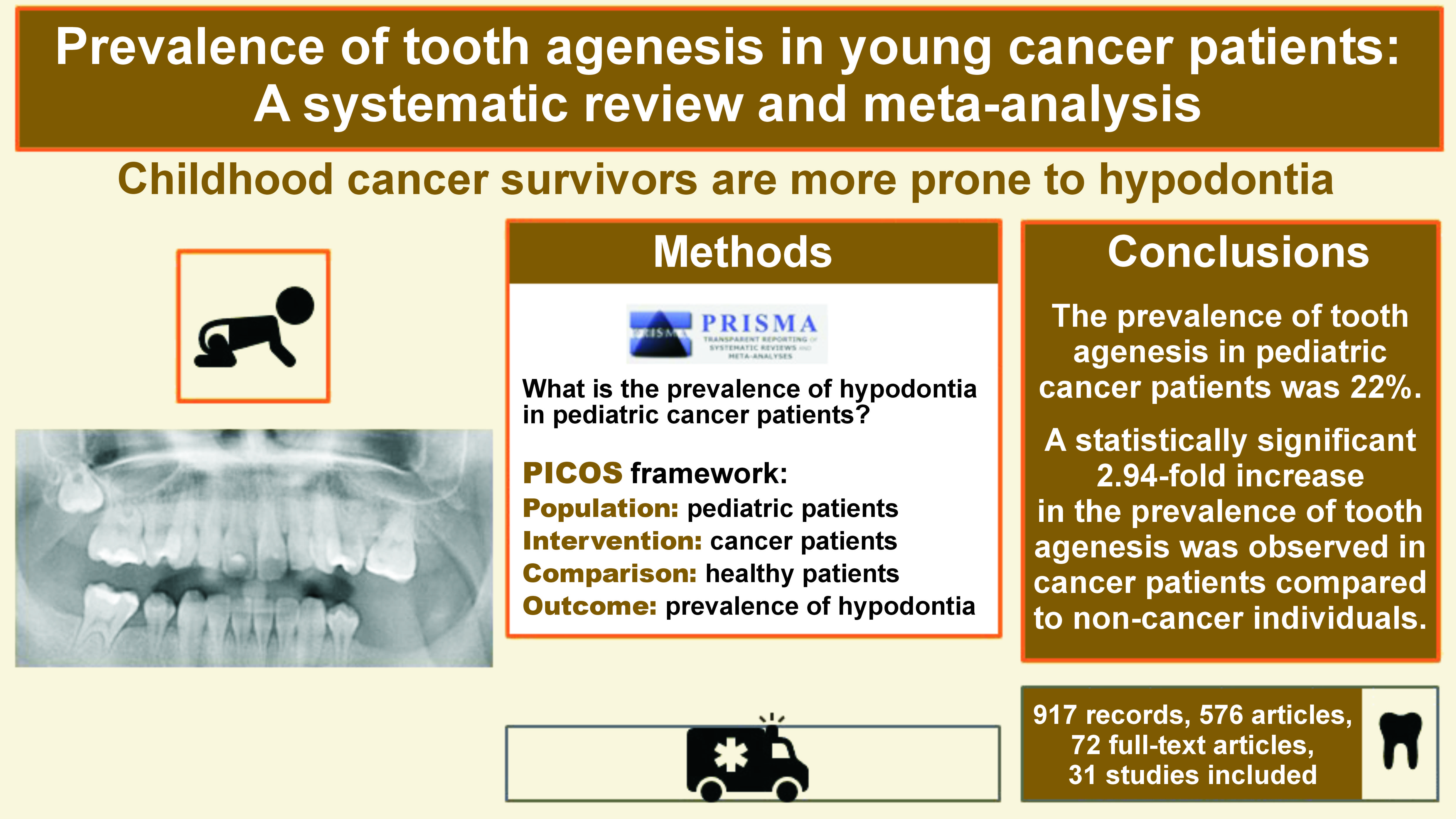
The present systematic review and meta-analysis was conducted according to the PRISMA (Preferred Reporting Items of Systematic Reviews and Meta-Analyses) guidelines in order to follow a uniform and transparent methodology.17 The study was registered with PROSPERO (registration No. CRD42022308068). The following PICOS (Population, Intervention, Comparison, Outcome, and Study design) framework was employed: Population – pediatric patients; Intervention – cancer patients; Comparison – healthy patients; Outcome – prevalence of hypodontia. The research question was: “What is the prevalence of hypodontia in pediatric cancer patients?”

Literature search

The systemic research strategy was implemented using multiple databases, namely PubMed®, Scopus, Web of Science, and Embase. The literature search was performed on February 21, 2023. The search strategy used in PubMed® and adapted in other database searches is presented in Table 1. After the search, all articles were imported into the Mendeley Desktop v. 1.17.11 software (Glyph & Cog, LLC, Petaluma, USA) to eliminate duplicates.

Study selection

The articles were imported into the Rayyan online tool,18 and the titles and abstracts were initially screened to identify studies that potentially met the following eligibility criteria: human experimental studies (cross-sectional and longitudinal, retrospective, and prospective) investigating the prevalence and patterns of tooth agenesis in pediatric patients with cancer; studies with at least 3 subjects with dental anomalies per group. Only manuscripts published in the English language were considered. Case series, case reports, pilot studies, and reviews were excluded from the analysis. The full texts of the articles were reviewed, and a systematic methodology was employed to label all the relevant information for the exclusion or inclusion of individual papers. The decision process was performed by 2 independent reviewers (PP and MLS). In the case of disagreement between the authors, the final decision was made through consultation with a third reviewer (CECS), a senior experienced researcher.

Data extraction

The relevant data from the included studies was extracted independently by 2 authors (PP and MLS) using a Microsoft Excel spreadsheet (Microsoft Corporation, Redmond, USA). In instances where information was incomplete or unclear, the authors of the included reports were contacted via e-mail for clarification. The following data was recorded for each included report: study design and sample size; age of participants during examination; age at diagnosis; cancer type; length of therapy; prevalence of hypodontia in cancer patients; and other dental anomalies.

Risk of bias

The risk of bias for all the included clinical trials was assessed by 2 independent reviewers (PP and MLS), and discrepancies were resolved by discussion and in consultation with a third reviewer (CECS). All included studies were evaluated using specific tools for each experimental design: the ROBINS-I (Risk Of Bias In Non-randomized Studies – of Interventions) for non-randomized clinical trials; the Newcastle–Ottawa Scale (NOS) for cohort studies; and the Joanna Briggs Institute (JBI) critical appraisal tool for cross-sectional and case–control studies.17

Statistical analysis

The data regarding the prevalence of tooth agenesis was pooled, and the risk difference with a 95% confidence interval (CI) was used as the effect size. Subsequently, the inverse variance method was selected to calculate the pooled effect. When data from the control patients was available, information regarding the prevalence of tooth agenesis in both cancer and non-cancer patients was used to gener­ate unadjusted odds ratios (ORs) and 95% CIs of the tooth agenesis for the cancer vs. non-cancer group. The hetero­geneity (I2) index and Cochran’s Q test were used to examine the heterogeneity between the studies. For the Cochran’s Q test, the p-value was significant at <0.05. All analyses were performed using the MedCalc® statistical software, v. 20.027 (MedCalc Software Ltd, Ostend, Belgium).

Results

Literature search

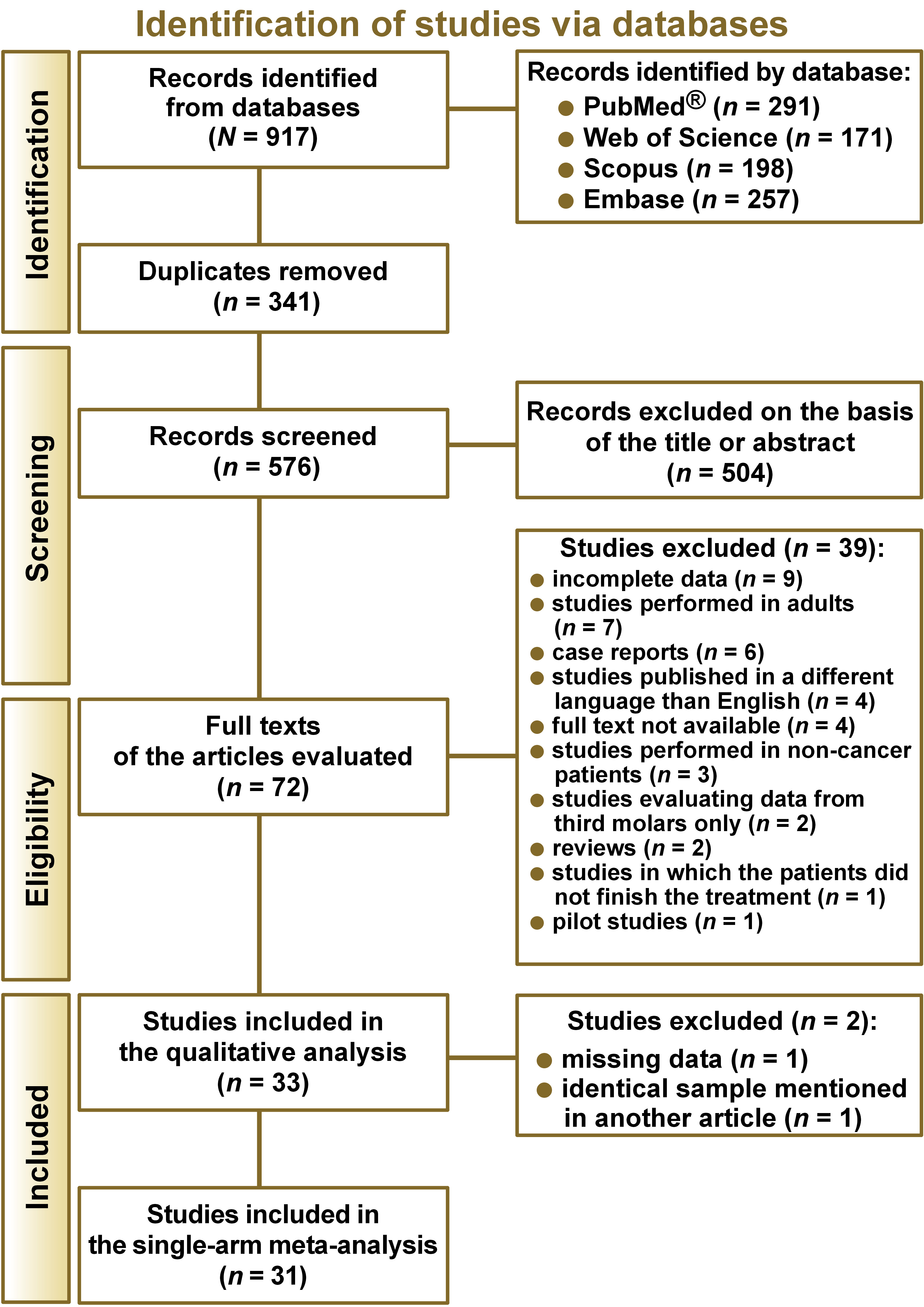
The literature search yielded a total of 917 records (Figure 1). After removing duplicates, 576 articles were screened, resulting in the exclusion of 504 papers based on the eligibility criteria. A total of 72 full-text articles were assessed for eligibility. Of these, 39 were not selected for the qualitative analysis. Nine of them did not present complete data, 7 were based on studies conducted exclusively on adults, 6 were case reports, 4 were published in a language other than English, 4 did not provide the full text, 3 were performed on non-cancer patients only, 2 evaluated data on third molars only, 2 were reviews, one of the studies included patients who did not complete the treatment, and 1 was a pilot study. A total of 33 studies were included in the qualitative analysis. However, 2 additional articles were excluded: one due to missing data; and the second one because it employed the same sample as another article. Finally, 31 studies were included in the single-arm meta-analysis.3, 6, 8, 9, 16, 19, 20, 21, 22, 23, 24, 25, 26, 27, 28, 29, 30, 31, 32, 33, 34, 35, 36, 37, 38, 39, 40, 41, 42, 43, 44, 45 Only 14 studies presented data for a control group and were included in the proportion meta-analysis.6, 12, 19, 20, 21, 22, 29, 30, 31, 32, 33, 34, 36, 44, 45

The characteristics of the included articles are summa­rized in Table 2. Several types of clinical studies were included, such as cross-sectional, cohort and case–control studies. In the investigated groups, the number of cancer patients ranged from 10 to 9,308. The subjects suffered from various forms of cancer, including solid tumors, leukemias and lymphomas. They were most often treated with CT alone; however, some patients also received RTX (including head and neck RTX), total body irradiation (TBI) and hematopoietic stem cell transplantation (HSCT). At the time of diagnosis, the majority of patients were under 10 years of age, with the youngest subject being 1 month old.

Various teeth were affected by agenesis. Most often missing teeth were second premolars, second molars and lower incisors.6, 8, 16, 19, 21, 22, 28, 30, 35, 36, 37, 43 Risk factors associated with a higher incidence of agenesis in cancer survivors were: younger age at diagnosis or treatment (1–7 years)3, 8, 16, 19, 20, 23, 27, 29, 32, 34, 35, 36, 37, 40, 43, 44, 46, 47, 48; use of multiple (>4) classes of chemotherapeutic agents, particularly alkyl­ating agents in high doses, and prolonged duration of therapy3, 16, 30, 37, 38; use of heavy metal compounds in CT2; RTX dosage greater than or equal to 2,200 cGy32, 44; head and neck radiation therapy (RTX)23, 35, 40; history of HSCT3, 37, 38, 43; and the presence of other dental anomalies.16

Risk of bias

For cross-sectional studies, the average quality score ranged between 4 and 6 (Table 3). The criteria that exhibited the highest failure rate pertained to the iden­tification of confounding factors. For cohort studies, the quality score ranged between 4 and 6 (Table 4). The studies under review failed to complete the inde­pendent blind assessment. Non-randomized clinical trials were catalogued as having a high risk of bias in domains of confounding and selection of participants into the study (Table 5). For case–control studies, the quality score ranged from 5 to 8. However, all studies failed to meet the criteria related to the identification and management of confounding factors (Table 6).

Meta-analysis

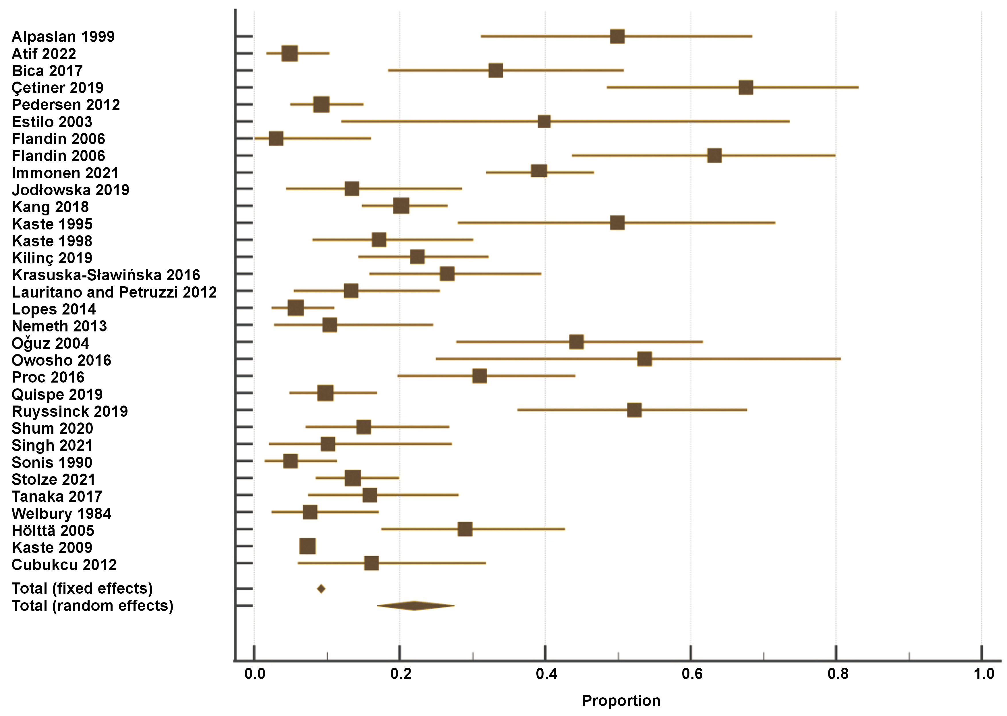
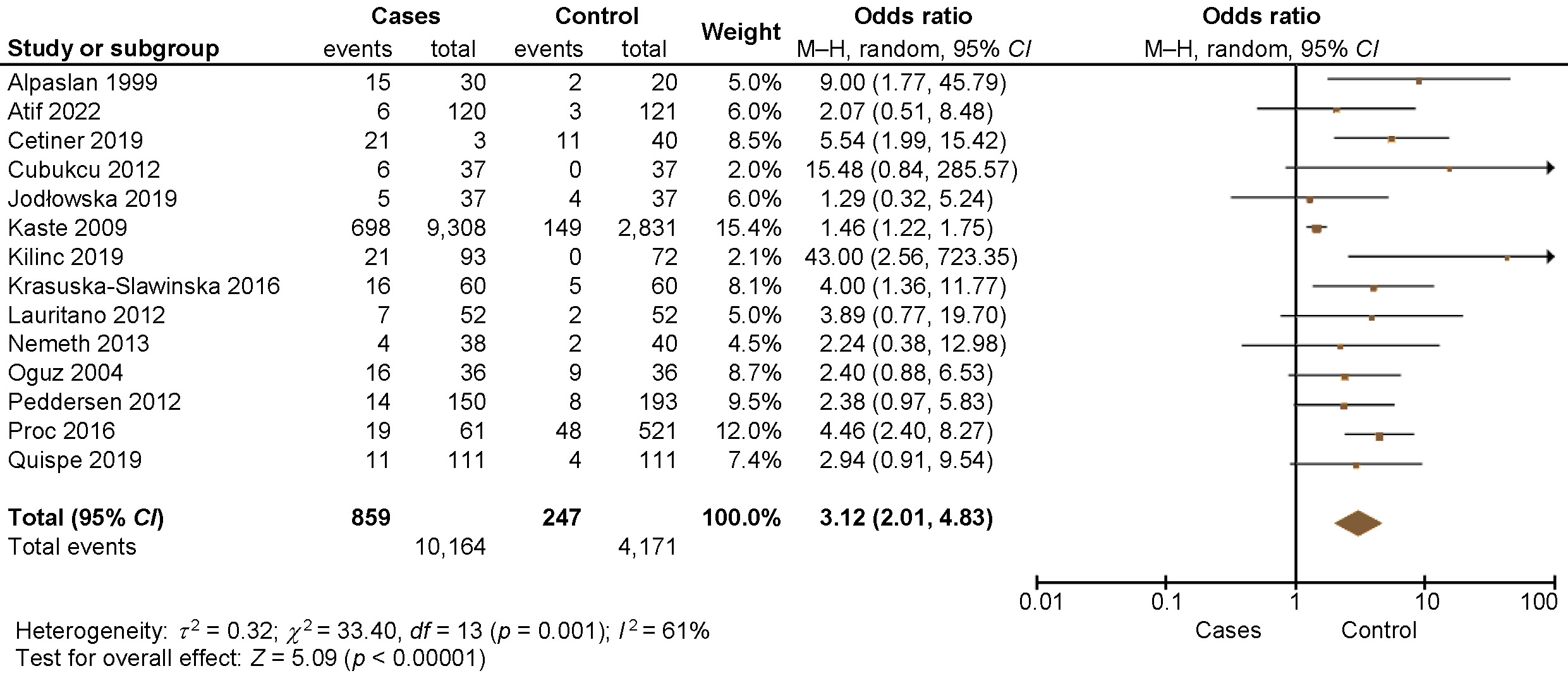
Figure 2 presents the results of the single-arm meta-analysis, which revealed that the prevalence of tooth agenesis in pediatric cancer patients was 22% (random effects model; 95% CI: 14–25%, p < 0.001). Pooled analyses of 15 unadjusted relative risk estimates demonstrated a statisti­cally significant 2.94-fold increase in the prevalence of tooth agenesis in cancer patients compared to non-cancer patients (unadjusted OR: 3.12; 95% CI: 2.01–4.83; p < 0.00001) (Figure 3). Dental abnormalities were found to be more common among cancer patients than in healthy controls in most of the reviewed studies.6, 19, 21, 30, 31, 33, 44, 45 All the details are provided in Table 2.

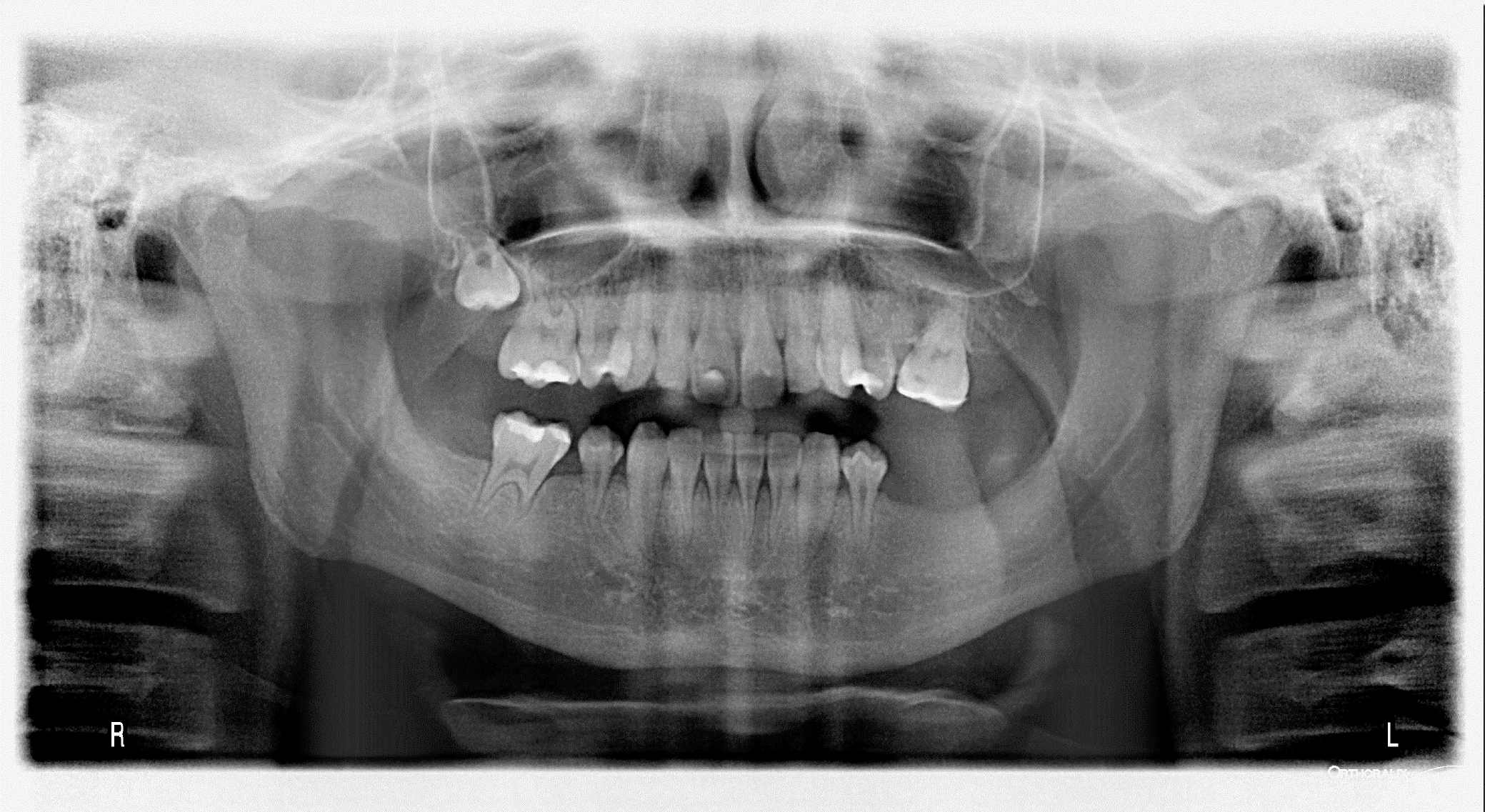
Figure 4 presents an exemplatory panoramic radiograph of a 15-year-old male patient diagnosed with neuroblastoma at the age of 3. The patient underwent a 21-month treatment regimen consisting of combination therapy, which included high-dose and conventional CT, bone marrow transplantation and RTX. The patient suffers from hypodontia, short roots of teeth and microdontia.

Discussion

This systematic review was aimed at assessing the prevalence of tooth agenesis in childhood cancer survivors and healthy individuals. The findings revealed that the occurence of hypodontia was higher in children who had undergone cancer treatment compared to their healthy peers. The null hypothesis stating that childhood cancer survivors and healthy individuals would have the same prevalence of tooth agenesis was rejected. The presence of defects depended on various factors, including both individual characteristics of the child and the applied treat­ment. According to the peer-reviewed articles, hypodontia was estimated to affect between 1.4% and 66.42% of cancer patients.3, 6, 8, 16, 19, 20, 21, 22, 23, 24, 25, 26, 27, 28, 29, 30, 31, 32, 33, 34, 35, 36, 37, 38, 39, 40, 41, 42, 43, 44, 45, 46 In the healthy group, the prevalence of hypodontia ranged from 0% to 25%.29, 34 The number of missing teeth in the cancer groups ranged from 6 to 69.6, 19, 27, 33, 35, 36, 46 It was also found that 15–85% of third molars were missing in cancer patients.19, 21

The teeth most frequently affected by agenesis were second premolars and second molars. In healthy individuals, the most often missing teeth were lateral incisors. The prevalence of specific groups of microdontic teeth depended on the time of the treatment and the conditions of the most active mineralization.13 A similar trend was observed with respect to the prevalence of hypodontia in particular tooth groups; however, the difference was not statistically significant. This phenomenon can be explained by the observation that, when exposed to particu­larly strong external factors, tooth buds undergo complete degradation, irrespective of their development stage.

Moreover, the overall dental development, as expressed by dental age, varied in cancer survivors and depended on the type of cancer and the implemented therapy.5 In the majority of cases, the dental age of cancer survivors was accelerated, predominantly due to premature closure of root apices. The dental age was significantly delayed in patients with familial adenomatous polyposis (FAP)-associated hepatoblastoma. However, the changes in dental age were independent of sex, age, or the duration of treatment.9, 49

Numerous factors can influence the occurrence of hypodontia, with the most prevalent one being the age of the patient at the time of diagnosis and therapy. The younger the age of the child at the time of diagnosis, the earlier the stage of tooth development and the greater the risk of more serious dental defects. The significant age limit varied in different publications, although it was consistently below 7 years of age.3, 8, 16, 19, 20, 23, 27, 29, 32, 33, 34, 35, 36, 37, 40, 43, 44, 46, 47 This is in line with the time of the most active development of tooth buds, which is considered to be the age between 6 and 8 years.6

The age at diagnosis correlated not only with the frequency but also with the severity of dental abnormalities. The patients in the youngest group presented with tooth agenesis or microdontia, while those in the oldest group demonstrated the most prevalent occurrence of abnormal root develop­ment.6, 20, 23, 26, 27, 29, 34 Additionally, the prevalence of combined disturbances was significantly lower in the youngest group compared to the other groups.2 The co-occurrence of differ­ent dental defects was frequently observed, as most of the cancer survivors suffered from more than one type of abnormality.5 Apart from hypodontia, the most frequently reported complications were microdontia, root deformation with premature apexification, enamel discoloration, and unerupted teeth. Patients with rhabdomyosarcoma of the head or neck who underwent treatment, including RTX, suffered from oral diseases, i.e., bony hypoplasia/facial asymmetry, trismus, velopharyngeal insufficiency, radiographically underdeveloped mandible, severe malocclusion, caries, hyposalivation/xerostomia, and gingivitis.23, 27, 35 On the other hand, factors like malocclusion, trauma, severe pain stimuli, parafunctional activities, and psychological elements, including stress, anxiety and depression can lead to temporomandibular disorders (TMD).50

It is worth noting that the dose, type and number of cytostatic drugs administered were identified as risk fac­tors for hypodontia and other dental defects. The use of more than 4 different chemotherapeutic agents and heavy metals has been identified as a significant risk factor for severe dental disturbances.2 Additionally, chemotherapeutic drugs such as vincristine, cyclophosphamide, doxorubicin, ifosfamide, etoposide, and cisplatin significantly increased the risk of tooth agenesis.5 Interestingly, it has been reported that equivalent doses of cyclophosphamide above 8,000 mg/m2 are associated with a higher number of teeth missing due to agenesis.6

Total body irradiation is performed in cancer patients to suppress the immune system and prevent the rejection of bone marrow transplantation (BMT).7 The side effects of TBI are most pronounced in terms of height and weight delay, while other complications of TBI include hypo­thyroidism, cataracts and a high incidence of secondary tumors.6 However, dental complications, such as tooth agenesis, were not found more frequently in the group of patients who had undergone TBI treatment.24, 43 As for patients treated with TBI, agenesis was more frequent in individuals receiving busulfan (63.2%) than in those treated with other chemotherapeutic agents (37.5%).7

On the other hand, some studies have documented a significantly higher prevalence of tooth agenesis in children treated with HSCT (similarly to BMT).2 The prevalence of agenesis and microdontia affecting at least 1 permanent tooth in cancer patients who had undergone HSCT treatment was much higher when compared to the controls. Moreover, 92.3% of children aged ≤3 years old at the time of HSCT treatment exhibited tooth agenesis.5 The condition manifested more prominently in certain tooth groups, including first and second premolars in the maxilla and mandible, as well as second molars in the mandible (all p-values <0.001).6

The relationship between the application of head and neck RTX and the occurrence of dental changes was also investigated.23, 35, 40 The radiation exposure of ≥20 Gy to the dentition was significantly associated with an increased risk of 1 or more dental abnormalities.6 After RTX, the frequency of dental changes reached from 80% up to 100% among children under 5 years of age.23, 40

Impaired tooth development constitutes a complication that arises subsequent to cancer treatment. Tumor-induced osteomalacia has been widely described in patients ranging in age from 9 months to 90 years, with a broad spectrum of tumor types. In adults, the primary concern is a decreased level of serum phosphate, while in children (aged <18 years), it is a low or improperly circulating concentration of 1,25-dihydroxyvitamin D.51 The 1,25-dihydroxyvitamin D, in turn, belongs to the group of interacting circulating hormones and their key receptors that regulate the state of calcium homeostasis.52 Calcium and phosphate play a key role in the mineralization of teeth and bones. Disturbances in the levels of these minerals during the developmental phase of an organism may partially account for the increased occurrence of dental defects in childhood cancer survivors.

Tooth agenesis is more prevalent among cancer survivors in comparison to healthy controls. There are several factors related to cancer and its treatment that contribute to the occurrence of agenesis. Given the high risk of complications in cancer patients, increased dental attention and care are required.

Conclusions

Patients who underwent childhood cancer treatment may experience dental complications more frequently compared to the general population. The dissemination of knowledge on this subject among clinicians is necessary to ensure the provision of specialized dental care to such patients, thereby facilitating their recovery and enhancing their quality of life.

Trial registration

The study was registered with PROSPERO (registration No. CRD42022308068).

Ethics approval and consent to participate

Not applicable.

Data availability

The datasets generated and/or analyzed during the current study are available from the corresponding author on reasonable request.

Consent for publication

Not applicable.

Use of AI and AI-assisted technologies

Not applicable.

Tables


Table 1. Search strategy

Search No.

Keywords

1

cancer patients OR pediatric cancer survivors OR pediatric stem cell transplantation OR blood transplantation OR marrow transplantation OR radiotherapy adverse effects OR chemotherapy adverse effect OR cervico-facial irradiation OR colorectal polyposis OR cancer

2

dental agenesis OR tooth agenesis OR hypodontia OR oligodontia OR anodontia

3

#1 and #2

The 3 searches were implemented across all databases (PubMed®, Scopus, Web of Science, and Embase).
Table 2. Characteristics of the included studies

Study

Type of study

Cancer patients, n

Cancer patients with hypodontia, n (%)

Age at examination

Age at cancer diagnosis

Cancer type

Follow-up period

Therapy received

Other dental anomalies evaluated

Alpaslan et al.19
1999

cross-sectional

30

15 (50.0)

4–15 years

ND

non-Hodgkin lymphoma (17); Hodgkin lymphoma (13)

17 months
(3–58 months)

CT

enamel discoloration; hypoplasia; unerupted teeth; premature apexification

Atif et al.20
2022

cross-sectional

120

6 (5.0)

>12 years

<8 years

acute lymphocytic leukemia (54); Hodgkin lymphoma (24); retinoblastoma (10); sarcomas (4); acute myeloid leukemia (9); medulloblastoma (1); Langerhans cell histiocytosis (2); PNET (3); non-Hodgkin lymphoma (13)

ND

CT

developmental defects of enamel

Bica et al.8
2017

cohort

36

12 (33.3)

10–12 years

1–6 years (n = 20);
7–12 years (
n = 16)

ALL

ND

CT

tooth eruption disorders (71%); hypoplasia (17%)

Çetiner et al.21
2019

cohort

53/31 who underwent dental examination

21 (39.6)

10 years ±4 months

ND

Hodgkin lymphoma (10); non-Hodgkin lymphoma (36); neuroblastoma (2); Wilms tumor (1); retinoblastoma (2); RMS (1); nasopharyngeal carcinoma (1)

1–5 years
(
M: 2 years ±4 months)

CT

enamel discoloration; enamel hypoplasia; unerupted teeth

Cubukcu et al.45
2012

case–control

37

6 (16.2)

ND

2.7 ±0.6 years

non-Hodgkin lymphoma (8); Wilms tumor (8); soft tissue sarcoma (4); medulloblastoma (3); optic glioma (1); neuroblastoma (4); Hodgkin lymphoma (3); retinoblastoma (2); Langerhans cell histiocytosis (2); other (hepatoblastoma and germ cell tumor) (2)

>5 years
(
M: 6.7 ±1.5 years)

CT and RTX

ND

Estilo et al.23
2003

cohort

10

4 (40.0)

10 years ±4 months

4.5 years (10 months–19.5 years)

RMS of the neck

12.2 years

CT and RTX

enamel defects; bony hypoplasia/facial asymmetry; trismus; velopharyngeal insufficiency; radiographically underdeveloped mandible; tooth agenesis; root agenesis; root stunting/tapering; arrested/incomplete root development

Flandin et al.24
2006

cohort

32 (TBI + CT);
30 (CT only)

TBI + CT: 1 (3.1);
CT: 19 (63.3)

TBI + CT: 181 (130–240) months;
CT: 198 (147–247) months

TBI + CT: M: 37 months;
CT:
M: 37 months

neuroblastoma

TBI + CT: 157 months
CT: 145 months

CT, RTX of the head or neck, TBI

ND

Hölttä et al.43
2005

cross-sectional

52

16 (31) patients without third molars;
77% (<3 years), 40% (3–5 years), and 0% (>5 years)

11.7 (4.7–25.7) years

10 years at the time of SCT

neuroblastoma; ALL; AML; chronic myeloid leukemia; myelodysplastic syndrome; severe aplastic anemia; RMS, yolk sac tumor

7.4 (1.0–20.6) years

CT, RTX of the head or neck, TBI

ND

Immonen et al.25
2021

cross-sectional

178

1.4–3.8%

ND

5.0 (2.5–16.8) years

ALL

6.3 (3.0–11.6) years

CT, RTX of the head or neck, TBI

ND

Jodłowska et al.26
2019

non-randomized clinical trial

37

5 (13.5)

<18 years

3 years and 2 months (range: 4 months–8 years and 6 months)

solid tumor (29); leukemia (8)

24–36 months

CT and RTX

ND

Kang et al.3
2018

cross-sectional

196

40 (20.4)

14.9 (4.6–33.9) years

4/7 years (0–16.4 years)

ALL (71); AML (7); lymphoma (23); brain tumor (22); sarcoma (18); abdomen (37); others (18)

6.9 (2.1–22.5) years

CT and RTX

enamel hypoplasia

Kaste et al.27
1995

cross-sectional

22

11 (50.0)

ND

5 years and 1 month

RMS of the head or neck

9 years and 5 months
(5–16 years)

CT and RTX

severe facial deformity; severe malocclusion; extensive caries

Kaste et al.28
1998

cross-sectional

52

9 (17.3)

ND

1.5 years (range: 3 days–7.2 years;
M: 1.9 years)

neuroblastoma

5.0 (1.9–19.3) years
(
M: 6.4 years)

CT and RTX

enamel hypoplasia (17%);
excessive caries (29%)

Kaste et al.44
2009

cross-sectional

8,522

698 (8.2)

ND

6.0 (0–20) years

leukemia (2,910 (34.2%)); CNS tumor (1,076 (12.6%)); Hodgkin lymphoma (1,086 (12.7%)); non-Hodgkin lymphoma (628 (7.4%)); Wilms tumor (794 (9.3%)); neuroblastoma (575 (6.8%)); soft tissue sarcoma (750 (8.8%)); bone cancer (702 (8.2%))

22.0 (15–34) years

CT and RTX

enamel hypoplasia; gingivitis; xerostomia

Kılınç et al.29
2019

case–control

93

21 (22.6)

9.54 ±1.25 years
(range: 8–13 years)

9 months–7 years

lymphoproliferative tumors; leukemia; lymphoma; Langerhans cell histiocytosis; solid tumors; neuroblastoma; renal tumor; soft tissue sarcoma; germ cell tumor; hepatic tumor; CNS tumor; retinoblastoma

5–8 years

CT and RTX

enamel defects (22 (23.7%))

Krasuska-Sławińska et al.30
2016

non-randomized clinical trial

60

16 (26.7)

11.81 ±3.87 years

5.9 ±4.0 years

Burkitt’s lymphoma (15.0%); nephroblastoma (13.0%); neuroblastoma (10.0%);
histiocytosis (8.3%); RMS (6.7%); Ewing sarcoma (6.7%); medulloblastoma (5.0%);
neurofibromatosis type I (5.0%); others (30.3%)

4.9 ±3.4 years

CT

root resorption (36 (60.0%));
enamel defects (53 (88.3%))

Lauritano and Petruzzi31
2012

non-randomized clinical trial

52

7 (13.5)

8–15 years

<15 months

ALL (39); AML (13)

60 ±24 months

CT and RTX

enamel hypoplasia (9 (17.3%))

Lopes et al.32
2006

cross-sectional

137

8 (5.8)

0–6; 6–12 years

5 years and 6 months

leukemia/lymphoma (61%); solid tumors (39%)

3–58 months
(
M: 17 months)

CT and RTX

microdontia (10 (7%)); taurodontism (19 (14%)); macrodontia (7 (5%)); blunted root (2 (2%)); tapered root (5 (4%))

Nemeth et al.33
2013

non-randomized clinical trial

38

4 (10.5) without third molars;
18 (47.4) with third molars

12.2 ±0.5 years

31 months–6 years;
M: 4.29 ±1.71 years

ND

6.9 ±2 years

CT, RTX of the head or neck, TBI

macrodontia (2–2.6%);
unerupted teeth (6–15.8%)

Oğuz et al.34
2004

non-randomized clinical trial

36

16 (44.4)

10.0 (4.2–17.6) years

7.1 years (range: 3.2–15 years)

non-Hodgkin lymphoma

2.6 (1–6.2) years

CT

enamel discoloration (24 (66.7%)); enamel defects (20 (55.6%)); unerupted teeth (7 (19.4%)); premature apexification (2 (5.6%))

Owosho et al.35
2016

cross-sectional

13

7 (53.8)

ND

5 years (range: 19
months–13 years)

RMS

9 (1–13) years

CT

facial asymmetry and jaw hypoplasia; trismus and hyposalivation/xerostomia; enamel malformation

Pedersen et al.22
2012

cohort

150

14 (9.3)

12–18 years

1–7 years

lymphomas and other reticuloendothelial neoplasms; CNS, intracranial and intraspinal neoplasms; sympathetic nervous system tumors; retinoblastoma; renal and hepatic neoplasms; bone and soft tissue sarcoma; gonadal neoplasms

ND

CT

ND

Proc et al.6
2016

case–control

61

19 (31.1)

5–18 years (56–213 months)

1–196 months

ALL; ANLL; B-cell non-Hodgkin lymphoma; PNET; germinal tumor; brain tumor; hepatoblastoma; neuroblastoma; RMS; Wilms tumor

4.9 years
(58.9 ±4.3 months)

CT and RTX

ND

Quispe et al.36
2019

case–control

111

11 (9.9)

M: 160.1 months

<192 months; M: 83.2 months

various

M: 18.3 months

CT, RTX of the head or neck, TBI

various but not significant

Ruyssinck et al.37
2019

case–control

42

51.3%

ND

<12 years

primitive neuroectodermal tumor (1); ALL (9); AML (2); juvenile myelomonocytic leukemia (2); neutropenia (severe, congenital) (1); neuroblastoma (9); Wilms tumor/nephroblastoma (2); anaplastic large cell lymphoma (1); juvenile metachromatic leukodystrophia (1); X-linked adrenoleukodystrophy (2); myelodysplastic syndrome (4); secondary myelodysplastic syndrome (1); chronic myeloid leukemia (2); aplastic anemia (2); thalassemia major (1); hemophagocytic lymphohistiocytosis (1); Burkitt’s lymphoma (1)

>1 year
(
M: 7 years)

CT and TBI

ND

Shum et al.38
2020

case–control

59

9 (15.3)

14–16 years; M: 14.9 ±0.80 years

<10 years; M: 4.1 ±2.9 years

various

ND

CT and RTX

ND

Singh et al.39
2021

case–control

29

3 (10.3)

37.3 (24.2–219.5) months

2.9 (0.8–14) years

neuroblastoma

ND

CT

hypocalcification of enamel; trismus

Sonis et al.40
1990

case–control

97

5 (5.2)

8 year and 1 month–16 years and 2 months

<10 years

ALL

5 years

CT and RTX

enamel hypoplasia

Stolze et al.16
2021

cross-sectional

154

21 (14.3)

32.4 (16.8–56.6) months

5.2 (0.3–16.1) years

hematological malignancy (111); brain tumor (7); solid tumor (36)

25.2 (15.9–48.8) months

CT, RTX of the head or neck, TBI

peg-shaped teeth; hypomineralization; persistent deciduous teeth

Tanaka et al.41
2017

cross-sectional

56

9 (16.1)

13.9 (4.6–32.7) years

1.9 (0.0–13.7) years

ALL (30 (53.6%)); AML (11 (19.6%)); juvenile myelomonocytic leukemia (1 (1.8%)); malignant lymphoma (4 (7.1%)); neuroblastoma (4 (7.1%)); Wilms tumor (2 (3.6%)); hepatoblastoma (1 (1.8%)); Langerhans cell histiocytosis (1 (1.8%));
retinoblastoma (1 (1.8%)); germinoma (1 (1.8%))

3 years from the completion of cancer treatment or 5 years from the time of the diagnosis

CT

enamel defects/hypoplasia (6 (10.7%))

Welbury et al.42
1984

cross-sectional

64

12 (18.8)

3–20 years

ND

leukemia (37); solid tumor (27)

ND

CT

hypoplastic teeth (36%)

ALL – acute lymphoblastic leukemia; ANLL – acute non-lymphoblastic leukemia; AML – acute myeloid leukemia; CNS – central nervous system; CT – chemotherapy; HSCT – hematopoietic stem cell transplantation; M – mean; ND – no data; PNET – primitive neuroectodermal tumor; RMS – rhabdomyosarcoma; RTX – radiotherapy; SCT – stem cell transplantation; TBI – total body irradiation.
Table 3. Assessment of the quality of studies using the Joanna Briggs Institute (JBI) critical appraisal tool for cross-sectional studies

Study

Q1

Q2

Q3

Q4

Q5

Q6

Q7

Q8

Score

Alpaslan et al.19
1999

N

Y

U

Y

N

N

Y

Y

4

Atif et al.20
2022

Y

Y

Y

Y

N

N

Y

Y

6

Hölttä et al.43
2005

Y

Y

Y

Y

N

N

Y

Y

6

Immonen et al.25
2021

Y

Y

U

Y

U

N

Y

Y

5

Kang et al.3
2018

U

U

Y

Y

N

N

Y

Y

4

Kaste et al.27
1995

Y

Y

Y

Y

N

N

Y

N

5

Kaste et al.28
1998

Y

Y

Y

Y

N

N

Y

N

5

Kaste et al.44
2009

Y

Y

Y

Y

N

N

Y

Y

6

Lopes et al.32
2006

Y

Y

Y

Y

N

N

Y

Y

6

Owosho et al.35
2016

N

Y

Y

Y

N

N

Y

N

4

Stolze et al.16
2021

Y

Y

Y

Y

N

N

Y

Y

6

Tanaka et al.41
2017

Y

Y

Y

Y

N

N

Y

Y

6

Welbury et al.42
1984

N

Y

Y

Y

N

N

Y

N

4

Y – yes; N – no; U – unclear.
Table 4. Assessment of the risk of bias using the Newcastle–Ottawa Scale (NOS) for cohort studies

Study

Selection

Comparability

Outcome

Total

representativeness of the exposed cohort

selection of the non-exposed cohort

ascertainment of exposure

outcome of interest not present at the start of the study

comparability of cohorts on the basis of the design or analysis

assessment of outcome

duration of follow-up

adequacy of follow-up

Bica et al.8
2017

1

1

1

1

0

0

0

0

4

Çetiner et al.21
2019

1

1

1

1

1

0

0

0

5

Estilo et al.23
2003

1

1

1

1

0

0

1

0

5

Flandin et al.24
2006

1

1

1

1

0

0

1

1

6

Pedersen et al.22
2012

1

1

1

1

1

0

0

0

5

Table 5. Assessment of the risk of bias using the ROBINS-I (Risk Of Bias In Non-randomized Studies – of Interventions) tool for non-randomized studies

Study

Bias due to confounding

Bias in selection of participants into the study

Bias in classification of interventions

Bias due to deviations from intended interventions

Bias due to missing data

Bias in measurement of outcomes

Bias in selection of the reported result

Jodłowska et al.26
2019

high

high

low

some concerns

low

low

low

Krasuska-Sławińska et al.30
2016

high

high

low

high

low

low

low

Lauritano and Petruzzi31
2012

high

high

low

high

low

low

low

Nemeth et al.33
2013

high

high

low

high

low

low

low

Oğuz et al.34
2004

high

high

low

some concerns

low

low

low

Table 6. Assessment of the quality of studies using the Joanna Briggs Institute (JBI) critical appraisal tool for case–control studies

Study

Q1

Q2

Q3

Q4

Q5

Q6

Q7

Q8

Q9

Q10

Overall score

Cubukcu et al.45
2012

Y

Y

Y

Y

Y

N

N

Y

Y

Y

8

Kılınç et al.29
2019

Y

Y

Y

Y

Y

N

N

Y

Y

Y

8

Proc et al.6
2016

Y

Y

U

Y

U

N

N

Y

Y

Y

6

Quispe et al.36
2019

Y

Y

Y

Y

Y

N

N

Y

Y

Y

8

Ruyssinck et al.37
2019

Y

Y

U

Y

U

N

N

Y

Y

Y

6

Shum et al.38
2020

Y

Y

U

Y

U

N

N

Y

Y

Y

6

Singh et al.39
2021

Y

Y

U

Y

U

N

N

Y

Y

N

5

Sonis et al.40
1990

Y

Y

U

Y

U

N

N

Y

N

Y

5

Figures


Fig. 1. Flowchart of the selection process
Fig. 2. Results of the single-arm meta-analysis presenting the prevalence of tooth agenesis in pediatric cancer patients
The study by Flandin et al. involved 2 types of patients: patients in the first group received radiation therapy, and the second group received chemotherapy.
Fig. 3. Forest plot for the prevalence of tooth agenesis in cancer patients compared to non-cancer patients
CI – confidence interval; df – degrees of freedom.
Fig. 4. Panoramic radiograph of a 15-year-old male patient diagnosed with neuroblastoma at the age of 3 years, who suffers from hypodontia, short roots of teeth and microdontia

References (52)

  1. Luongo C, Randazzo E, Iughetti L, et al. Cardiometabolic risk in childhood cancer survivors. Minerva Pediatr (Torino). 2021;73(6):588–605. doi:10.23736/S2724-5276.21.06544-7
  2. Dieffenbach BV, Liu Q, Murphy AJ, et al. Late-onset kidney failure in survivors of childhood cancer: A report from the Childhood Cancer Survivor Study. Eur J Cancer. 2021;155:216–226. doi:10.1016/j.ejca.2021.06.050
  3. Kang CM, Hahn SM, Kim HS, et al. Clinical risk factors influencing dental developmental disturbances in childhood cancer survivors. Cancer Res Treat. 2018;50(3):926–935. doi:10.4143/crt.2017.296
  4. van Iersel L, Mulder RL, Denzer C, et al. Hypothalamic-pituitary and other endocrine surveillance among childhood cancer survivors. Endocr Rev. 2022;43(5):794–823. doi:10.1210/endrev/bnab040
  5. Strebel S, Waespe N, Kuehni CE. Hearing loss in childhood cancer survivors. Lancet Child Adolesc Health. 2021;5(5):e17. doi:10.1016/S2352-4642(21)00099-7
  6. Proc P, Szczepańska J, Skiba A, Zubowska M, Fendler W, Młynarski W. Dental anomalies as late adverse effect among young children treated for cancer. Cancer Res Treat. 2016;48(2):658–667. doi:10.4143/crt.2015.193
  7. Patni T, Lee CT, Li Y, et al. Factors for poor oral health in long-term childhood cancer survivors. BMC Oral Health. 2023;23(1):73. doi:10.1186/s12903-023-02762-0
  8. Bica C, Chincesan M, Esian D, et al. Dental development in children after chemotherapy. Rev Chim. 2017;68(6):1397–1400. doi:10.37358/rc.17.6.5681
  9. Proc P, Szczepańska J, Zubowska M, Zalewska-Szewczyk B, Młynarski W. The broad variability in dental age observed among childhood survivors is cancer specific. Cancer Res Treat. 2021;53(1):252–260. doi:10.4143/crt.2020.275
  10. Feghali S, Vi-Fane B, Picard A, Kadlub N. Dental and orthodontic follow-up in nevoid basal cell carcinoma syndrome patient with odontogenic keratocystic tumors. J Stomatol Oral Maxillofac Surg. 2022;123(3):e57–e61. doi:10.1016/j.jormas.2021.07.009
  11. Qin W, Wan QQ, Ma YX, et al. Manifestation and mechanisms of abnormal mineralization in teeth. ACS Biomater Sci Eng. 2023;9(4):1733–1756. doi:10.1021/acsbiomaterials.1c00592
  12. Jodłowska A, Postek-Stefańska L. Systemic anticancer therapy details and dental adverse effects in children. Int J Environ Res Public Health. 2022;19(11):6936. doi:10.3390/ijerph19116936
  13. Rabassa-Blanco J, Brunet-Llobet L, Marcote-Sinclair P, Balsells-Mejía S, Correa-Llano MG, Miranda-Rius J. Prevalence of, and risk factors for, dental sequelae in adolescents who underwent cancer therapy during childhood. Oral Dis. 2024;30(2):604–614. doi:10.1111/odi.14317
  14. Halperson E, Matalon V, Goldstein G, et al. The prevalence of dental developmental anomalies among childhood cancer survivors according to types of anticancer treatment. Sci Rep. 2022;12(1):4485. doi:10.1038/s41598-022-08266-1
  15. Jensen JM, Skakkebæk A, Gaustadness M, et al. Familial colorectal cancer and tooth agenesis caused by an AXIN2 variant: How do we detect families with rare cancer predisposition syndromes? Fam Cancer. 2022;21(3):325–332. doi:10.1007/s10689-021-00280-y
  16. Stolze J, Vlaanderen KCE, Holtbach FCED, et al. Long-term effects of childhood cancer treatment on dentition and oral health: A dentist survey study from the DCCSS LATER 2 Study. Cancers (Basel). 2021;13(21):5264. doi:10.3390/cancers13215264
  17. Page MJ, McKenzie JE, Bossuyt PM, et al. The PRISMA 2020 statement: An updated guideline for reporting systematic reviews. Syst Rev. 2021;10(1):89. doi:10.1186/s13643-021-01626-4
  18. Ouzzani M, Hammady H, Fedorowicz Z, Elmagarmid A. Rayyan – a web and mobile app for systematic reviews. Syst Rev. 2016;5(1):210. doi:10.1186/s13643-016-0384-4
  19. Alpaslan G, Alpaslan C, Gögen H, Oğuz A, Cetiner S, Karadeniz C. Disturbances in oral and dental structures in patients with pediatric lymphoma after chemotherapy: A preliminary report. Oral Surg Oral Med Oral Pathol Oral Radiol Endod. 1999;87(3):317–321. doi:10.1016/s1079-2104(99)70215-5
  20. Atif M, Mathur VP, Tewari N, Bansal K, Rahul M, Bakhshi S. Long-term effect of anticancer therapy on dentition in childhood cancer survivors: An observational, cross-sectional study. Indian J Pediatr. 2022;89(4):327–332. doi:10.1007/s12098-021-03818-1
  21. Çetiner D, Çetiner S, Uraz A, et al. Oral and dental alterations and growth disruption following chemotherapy in long-term survivors of childhood malignancies. Support Care Cancer. 2019;27(5):1891–1899. doi:10.1007/s00520-018-4454-0
  22. Pedersen LB, Clausen N, Schrøder H, Schmidt M, Poulsen S. Microdontia and hypodontia of premolars and permanent molars in childhood cancer survivors after chemotherapy. Int J Paediatr Dent. 2012;22(4):239–243. doi:10.1111/j.1365-263X.2011.01199.x
  23. Estilo CL, Huryn JM, Kraus DH, et al. Effects of therapy on dentofacial development in long-term survivors of head and neck rhabdomyosarcoma: The memorial sloan-kettering cancer center experience. J Pediatr Hematol Oncol. 2003;25(3):215–222. doi:10.1097/00043426-200303000-00007
  24. Flandin I, Hartmann O, Michon J, et al. Impact of TBI on late effects in children treated by megatherapy for Stage IV neuroblastoma. A study of the French Society of Pediatric Oncology. Int J Radiat Oncol Biol Phys. 2006;64(5):1424–1431. doi:10.1016/j.ijrobp.2005.10.020
  25. Immonen E, Nikkilä A, Peltomäki T, Aine L, Lohi O. Late adverse effects of childhood acute lymphoblastic leukemia treatment on developing dentition. Pediatr Blood Cancer. 2021;68(9):e29200. doi:10.1002/pbc.29200
  26. Jodłowska A, Sobol-Milejska G, Postek-Stefańska L. A critical look at prevalence assessment of dental abnormalities after chemotherapy. Clinical research. J Stoma. 2019;72(3):95–105. doi:10.5114/jos.2019.87522
  27. Kaste SC, Hopkins KP, Bowman LC. Dental abnormalities in long-term survivors of head and neck rhabdomyosarcoma. Med Pediatr Oncol. 1995;25(2):96–101. doi:10.1002/mpo.2950250209
  28. Kaste SC, Hopkins KP, Bowman LC, Santana VM. Dental abnormalities in children treated for neuroblastoma. Med Pediatr Oncol. 1998;30(1):22–27. doi:10.1002/(sici)1096-911x(199801)30:1<22::aid-mpo8>3.0.co;2-2
  29. Kılınç G, Bulut G, Ertuğrul F, et al. Long-term dental anomalies after pediatric cancer treatment in children. Turk J Haematol. 2019;36(3):155–161. doi:10.4274/tjh.galenos.2018.2018.0248
  30. Krasuska-Sławińska E, Brożyna A, Dembowska-Bagińska B, Olczak-Kowalczyk D. Antineoplastic chemotherapy and congenital tooth abnormalities in children and adolescents. Contemp Oncol (Pozn). 2016;20(5):394–401. doi:10.5114/wo.2016.64602
  31. Lauritano D, Petruzzi M. Decayed, missing and filled teeth index and dental anomalies in long-term survivors leukaemic children: A prospective controlled study. Med Oral Patol Oral Cir Bucal. 2012;17(6):e977–e980. doi:10.4317/medoral.17955
  32. Lopes NNF, Petrilli AS, Caran EMM, França CM, Chilvarquer I, Lederman H. Dental abnormalities in children submitted to antineoplastic therapy. J Dent Child (Chic). 2006;73(3):140–145. PMID:17367030.
  33. Nemeth O, Hermann P, Kivovics P, Garami M. Long-term effects of chemotherapy on dental status of children cancer survivors. Pediatr Hematol Oncol. 2013;30(3):208–215. doi:10.3109/08880018.2013.763391
  34. Oğuz A, Cetiner S, Karadeniz C, Alpaslan G, Alpaslan C, Pinarli G. Long-term effects of chemotherapy on orodental structures in children with non-Hodgkin’s lymphoma. Eur J Oral Sci. 2004;112(1):8–11. doi:10.1111/j.0909-8836.2004.00094.x
  35. Owosho AA, Brady P, Wolden SL, et al. Long-term effect of chemotherapy-intensity-modulated radiation therapy (chemo-IMRT) on dentofacial development in head and neck rhabdomyosarcoma patients. Pediatr Hematol Oncol. 2016;33(6):383–392. doi:10.1080/08880018.2016.1219797
  36. Quispe RA, Rodrigues ACC, Buaes AMG, Capelozza ALA, Rubira CMF, Santos PSS. A case–control study of dental abnormalities and dental maturity in childhood cancer survivors. Oral Surg Oral Med Oral Pathol Oral Radiol. 2019;128(5):498–507.e3. doi:10.1016/j.oooo.2019.07.005
  37. Ruyssinck L, Toulouse K, Bordon Cueto de Braem V, Cauwels R, Dhooge C. Impact of hematopoietic stem cell transplantation on dental development. Biol Blood Marrow Transplant. 2019;25(1):107–113. doi:10.1016/j.bbmt.2018.08.027
  38. Shum M, Mahoney E, Naysmith K, et al. Associations between childhood cancer treatment and tooth agenesis. N Z Med J. 2020;133(1523):41–54. PMID:33032302.
  39. Singh A, Modak S, Solano AK, et al. Mandibular metastases in neuroblastoma: Outcomes and dental sequelae. Pediatr Blood Cancer. 2021;68(4):e28918. doi:10.1002/pbc.28918
  40. Sonis AL, Tarbell N, Valachovic RW, Gelber R, Schwenn M, Sallan S. Dentofacial development in long-term survivors of acute lymphoblastic leukemia. A comparison of three treatment modalities. Cancer. 1990;66(12):2645–2652. doi:10.1002/1097-0142(19901215)66:12<2645::aid-cncr2820661230>3.0.co;2-s
  41. Tanaka M, Kamata T, Yanagisawa R, et al. Increasing risk of disturbed root development in permanent teeth in childhood cancer survivors undergoing cancer treatment at older age. J Pediatr Hematol Oncol. 2017;39(3):e150–e154. doi:10.1097/MPH.0000000000000788
  42. Welbury RR, Craft AW, Murray JJ, Kernahan J. Dental health of survivors of malignant disease. Arch Dis Child. 1984;59(12):1186–1187. doi:10.1136/adc.59.12.1186
  43. Hölttä P, Alaluusua S, Saarinen-Pihkala UM, Peltola J, Hovi L. Agenesis and microdontia of permanent teeth as late adverse effects after stem cell transplantation in young children. Cancer. 2005;103(1):181–190. doi:10.1002/cncr.20762
  44. Kaste SC, Goodman P, Leisenring W, et al. Impact of radiation and chemotherapy on risk of dental abnormalities: A report from the Childhood Cancer Survivor Study. Cancer. 2009;115(24):5817–5827. doi:10.1002/cncr.24670
  45. Cubukcu CE, Sevinir B, Ercan I. Disturbed dental development of permanent teeth in children with solid tumors and lymphomas. Pediatr Blood Cancer. 2012;58(1):80–84. doi:10.1002/pbc.22902
  46. van der Pas-van Voskuilen IGM, Veerkamp JSJ, Raber-Durlacher JE, et al. Long-term adverse effects of hematopoietic stem cell transplantation on dental development in children. Support Care Cancer. 2009;17(9):1169–1175. doi:10.1007/s00520-008-0567-1
  47. Aarnoutse R, de Vos-Geelen JMPGM, Penders J, et al. Study protocol on the role of intestinal microbiota in colorectal cancer treatment: A pathway to personalized medicine 2.0. Int J Colorectal Dis. 2017;32(7):1077–1084. doi:10.1007/s00384-017-2819-3
  48. Tanem KE, Stensvold E, Wilberg P, Skaare AB, Brandal P, Herlofson BB. Oral and dental late effects in long-term survivors of childhood embryonal brain tumors. Support Care Cancer. 2022;30(12):10233–10241. doi:10.1007/s00520-022-07405-8
  49. Hölttä P, Hovi L, Saarinen-Pihkala UM, Peltola J, Alaluusua S. Disturbed root development of permanent teeth after pediatric stem cell transplantation. Dental root development after SCT. Cancer. 2005;103(7):1484–1493. doi:10.1002/cncr.20967
  50. Minervini G, Franco R, Marrapodi MM, et al. Correlation between temporomandibular disorders (TMD) and posture evaluated through the Diagnostic Criteria for Temporomandibular Disorders (DC/TMD): A systematic review with meta-analysis. J Clin Med. 2023;12(7):2652. doi:10.3390/jcm12072652
  51. Bosman A, Palermo A, Vanderhulst J, et al. Tumor-induced osteomalacia: A systematic clinical review of 895 cases. Calcif Tissue Int. 2022;111(4):367–379. doi:10.1007/s00223-022-01005-8
  52. Matikainen N, Pekkarinen T, Ryhänen EM, Schalin-Jäntti C. Physiology of calcium homeostasis: An overview. Endocrinol Metab Clin North Am. 2021;50(4):575–590. doi:10.1016/j.ecl.2021.07.005